Denis J. Marcellin-Little, DEDV, DipACVS, DipECVS North Carolina State University, USA
Denis J. Marcellin-Little is Assistant Professor, Department of Orthopedic Surgery, Department of Home and exotic species Animals of the Veterinary College of the University of North Carolina, USA
MAIN PROVISIONS
- Humerus fractures are common in dogs and account for 10% of all limb fractures. They are usually the result of traffic accidents, falls, gunshot wounds and minor injuries.
- There are three classic types of humerus fractures: approximately 20% of all fractures are physeal fractures in young dogs; 50% - diaphyseal fractures observed with more severe injuries (half of these fractures are comminuted); about 20% are condylar fractures in adult dogs, observed as a result of insufficient ossification of the condyle of the humerus.
- In most cases, humerus fractures require surgical treatment. Condylar fractures are well cured with a screw that tightens the bone fragments. Non-displaced diaphyseal fractures can be repaired by inserting a rod into the bone and fixing the bone fragments with wire; displaced fractures of this type can be repaired by placing plates on bone fragments, internal fixing nails or external fixators.
- Early postoperative physiotherapy is helpful to help restore elbow mobility and limb function. From the author's point of view, physiotherapy plays essential role in the success of the treatment of humerus fractures in dogs.
INTRODUCTION
Humerus fractures are common in dogs. In a recent epidemiological study, out of 30,140 fractures in dogs, the humerus accounts for 34% of all forelimb fractures and 10% of total fractures (1). Fractures of this kind occur in dogs of all breeds and ages. Fractures can occur in the proximal part of the humerus (in the head of the bone, in the area of the large tubercle), in the tubular part of the bone (diaphysis), in the distal part (in the region of the condyle, middle or lateral supracondylar zone). Sometimes there are combined fractures in several of these areas at once.
This article discusses the etiology, manifestations, clinical symptoms, diagnosis and treatment of humerus fractures in dogs. Clinical data are based on previous work (2-7) and on a retrospective analysis of 243 controlled cases of humerus fractures in dogs admitted to the veterinary teaching hospital at the NC State University College of Veterinary Medicine between September 1983 and November 1995.
All typical fractures of the humerus can be divided into 3 types. First, there are condylar bone fractures that occur with minor trauma, usually from a fall from a height of three feet or less, in young, prepubescent dogs (5). Secondly, fractures, mainly of the shaft of the humerus, resulting from road traffic accidents in adult dogs. Third, adult dogs of some breeds are prone to condylar fractures of the humerus with little or no visible trauma. These fractures are associated with incomplete ossification of the humeral condyle region in these breeds (8). The following breeds are predisposed to such fractures: cocker spaniels (8-11); springer spaniels (4,10,12); English spaniels (8.9); Cavalier King Charles Spaniels (4.12); Labrador Retrievers (13) and, less commonly, mestizos (8).
Manifestations
Humeral fractures occur in dogs of all breeds. In this study, such fractures were presented in dogs of 52 breeds. They were most commonly seen in English Spaniels, Great Danes and Labrador Retrievers (Table 1). Miniature poodles are the least susceptible to this type of injury (confidence interval 0.01-0.95). However, according to other authors who analyzed the statistics of 189 condylar fractures of the humerus in dogs of 58 breeds, English and French bulldogs, Yorkshire terriers and miniature schnaupers were the most susceptible to this injury, and there was no significant effect of breed size on the frequency of such fractures (7 ).
Three quarters of all condylar fractures of the humerus in puppies occur between 3 and 5 months of age (7). The frequency of fractures resulting from traffic accidents did not depend on the age of the animals. On average, the age of the dogs that suffered a fracture of the humerus in such incidents was 4 years. In the group of cocker spaniels with fractures due to incomplete ossification of the condyle of the humerus average age was 6 years (interval - from 2 to 11 years).
There was no correlation between the frequency of condylar fractures in puppies and their gender. However, based on an analysis of the entire sample of dogs treated at the NC State Veterinary Clinic, the risk of humerus fracture is highest in healthy males (CI 1.87 to 3.16). In neutered males, this risk is significantly lower (CI 0.4–0.94), and the risk of fracture is even lower in bitches (CI 0.24–0.5). In Cocker Spaniels, males (both normal and neutered) compared with females had a 3-5 times higher incidence of fractures associated with insufficient ossification of the condyle of the humerus.
The weight of the animals had almost no effect on the incidence of fractures of the humerus. Although one study has shown that heavier Cocker Spaniels are more likely to develop fractures due to incomplete ossification of the humeral condyle (8), this has not been confirmed by other authors (14).
Table 1
Distribution of those treated at the University of North Carolina in 1983-1995. dogs by breed and predisposition to such fractures
| Breed | Number of humerus fractures * | Quantity
hospitalization forged dogs |
min./max. ** | 95% confidence interval |
| English spaniel |
7 | 170 | 6,31 | 2.75 to 25.6 |
| Great Dane | 7 | 387 | 2,83 | 2.69 to 14.1 |
| cocker spaniel | 32 | 1,941 | 2,68 | 1.22 to 6.24 |
| Labrador Retriever | 32 | 2,628 | 1,94 | 1.31 to 2.86 |
| Metis | 49 | 7,425 | nd | nd |
| German Shepherd | 14 | 1,388 | nd | nd |
| Golden retriever |
10 | 1,962 | nd | nd |
| Boston Terrier | 6 | 371 | nd | nd |
| Beagle | 5 | 392 | nd | nd |
| chow chow | 5 | 395 | nd | nd |
| Rottweiler | 5 | 696 | nd | nd |
| Yorkshire Terrier Other breeds | | 5 | 458 | nd | nd |
| (<5 собак) Всего | 66 | 18,076 | nd | nd |

Picture 1. Mediolateral and craniocaudal radiographs of the humerus in a 15 week old male beagle.
(a) Lateral fracture of the condyle (area Salter IV) caused by an unknown injury. The fracture was fastened with a bone screw 3.5 mm in diameter, tightening the bone fragments, a washer and a Kirschner wire 1.6 mm in diameter.
(b) 6 weeks after surgical bonding of the fracture. The fracture is healed.
Etiology
In puppies, condylar fractures of the humerus are most common. According to the work (15), of all the studied cases of such fractures in puppies, 78% were due to physeal fractures in the Salter region. IV (Figure 1a), 16% were fractures in Salter II, 3% - in Salter I (Figure 2a) and 3% fractures in Salter III. Fractures also occur in the proximal epiphysis of the humerus, usually in the Salter areas. I and salter II. The prevalence of such fractures is determined in part by the relative softness of the physeal cartilage compared to the surrounding bone and soft tissues. Perhaps the asymmetry of the load of the radial head on the lateral side of the condyle of the humerus also plays a role. According to one of the messages (2), 20% of all humerus fractures (20/107) are physeal.
It is assumed that fractures of the humerus in falls and traffic accidents are associated with its strong load. The condyle of the humerus fractures in falls due to severe overexertion and twisting of the limb along with sprain of the elbow joint resulting in contact of the olecranon with the caudal part of the supracondylar zone of the humerus. The olecranon plays the role of a wedge driven between the medial and lateral parts of the condyle of the humerus (5). Fifty-eight percent of all dogs with shoulder fractures treated at NC State Hospital had serious injuries: 38% were victims of traffic accidents, 11% fell more than 3 feet, and 9% were shot.
In dogs with incomplete ossification of the condyle, a fibrous plate in the form of a cartilaginous area separating the medial and lateral parts of the condyle of the humerus, which normally disappears in dogs at 10 months of age, persists throughout life (8). The presence of this plate reduces the mechanical strength of the condyle, which makes such animals prone to fractures of the humerus. (Picture 3).

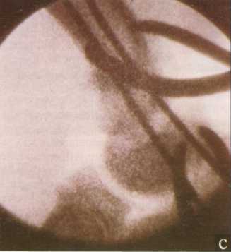
Figure 2. (a) Craniocaudal radiograph of the elbow of a 4-month-old mestizo male (German Shepherd/Chow-Chow mix). Visible fracture in the area Salter I of the humerus with severe soft tissue edema.
(b) Caudal surgical approach to the fracture, including tenotomy of the triceps tendon. Fracture aligned and crossed two Kirschner wires with a diameter of 1.6 mm, used as intraosseous wires.


(c) X-ray of the humerus taken with the help of a portable X-ray machine to assess the correct placement of the pins during surgery.
(d) Mediolateral and craniocaudal radiographs taken 4 weeks after surgery. The fracture is healed.
The pathogenesis of incomplete ossification of the condyle may be the same that results in fragmentation of the medial coronoid process of the humerus, also common in these dogs (16). Of NC State hospital-treated dogs with humerus fractures, 22% had incomplete condylar ossification.
Rarely, unusual cases of fractures of the humerus are observed. In one dog seen at the North Carolina State University hospital, this fracture was caused by fibroblastic osteosarcoma. The literature mentions a case of a secondary condylar fracture in a dog with a bone cyst aneurysm (17).
Clinical symptoms
Of all dogs with humerus fractures seen at the NC State Hospital, 84% were holding the injured limb in the air; 4% lightly touched the ground with her fingers; 4% stepped on a limb but limped heavily and 2% had intermittent claudication. In the remaining 5% of dogs, the degree of lameness could not be clinically determined. Dogs with incomplete ossification of the condyle often suffered a chronic fracture in this area of the bone with little displacement of the fragments. Such dogs, although lame, are able to step on an injured limb. In 31% of dogs with fractures of the process of the humerus due to traffic accidents, chest injuries (pneumothorax, pulmonary contusions) were also observed. However, radial nerve injury or avulsion of the brachial plexus in humerus fractures is uncommon.
Diagnostics
Humerus fractures are established by clinical examination and confirmed by radiography of the mediolateral and craniocaudal sections of the humerus.
Figure 3 Mediolateral and craniocaudal radiographs of the elbow joint of an 11-year-old castrated male Cocker Spaniel.

(a) Lateral condylar fracture of the humerus on the right, caused by an unknown injury.

(b) The fracture was fastened with a bone screw with a diameter of 4.5 mm with compression of the bone fragments.

(c) On the left side, with a cranio-caudal projection, only a small notch of 2 mm is visible in the region of the apex of the convexity of the condyle.

(d) On the scan of the left elbow joint, the notch is visible in the caudal zone of the condyle. Sclerosis of the surrounding bone tissue is also noticeable.

(e) Six months later, due to an unknown cause, a slightly displaced lateral-condylar fracture occurred in the left condyle.
In growing dogs, contralateral radiography can also be performed for control. In dogs under 1 year of age, 75% of condylar fractures are lateral, 16% are intracondylar, and 9% are midcondylar. (Figure 4). However, in dogs with incomplete condylar ossification, only 34% of fractures are lateral, 14% are midcondylar, and 51% are intracondylar (8). Although it is believed that 93% of dogs with incomplete condyle ossification have unilateral lesions, in our clinical practice humeral lesions in these dogs have always been bilateral, as evidenced by the presence of a radiolucent line in the opposite humeral condyle from the fracture site. (Figures 3c, d). In 25% of animals with incomplete ossification, obvious fractures were detected in the opposite condyle. (Figure Ze).
With optimal ossification of the condyle, bilateral fractures in this part of the humerus are rare in dogs. In transport injuries, the fracture is usually localized in the diaphysis of the humerus, and in 50% of cases these fractures are comminuted, in 25% with displacement of bone fragments, and in 25% they are oblique (2).
Treatment
Before surgery, dogs should be placed in a cage to limit mobility. Concomitant systemic diseases require immediate detection. A splint splint can be placed around the fracture site, but its usefulness is debated. Soft dressings with pads should be avoided, because. they exert pressure on the fracture site without limiting the mobility of the bone fragments. Sometimes such a bandage can be a fulcrum, due to which the parts of the broken bone are displaced relative to each other. The distribution of dogs treated at the North Carolina State University clinic by treatment for humerus fracture was as follows: 170 animals (70%) used internal fixation of bone fragments; 34 (14%) dogs had a combination of internal and external fixation; in 7 (3%) only external fixation; 2 dogs underwent arthrodesis of the elbow joint; 6 dogs were treated conservatively; in 9, the limb had to be amputated; 8 dogs were euthanized and 7 had treatment interrupted.

Figure 4 Condylar fractures of the humerus:(a) intercondylar;(b) lateral-condylar;(with) mid-condylar;(d) supracondylar.
Conservative treatment
Conservative treatment of fractures of the humerus is rarely used. This approach is possible when the fracture is undisplaced, does not involve the joint, and occurs in a young, growing dog. In conservative treatment of a fracture, the injured limb is fixed with a lateral (spiral) splint or Velpeau bandage for 2-3 weeks. The purpose of this fixation is to limit mobility at the fracture site.
Surgical recovery
Most humerus fractures require surgical treatment. Such treatment is indicated if the fracture involves the joint, is unstable, is characterized by difficulty in preventing displacement of bone fragments, or the likelihood of loss of mobility of the elbow joint due to prolonged immobilization and lack of load. Surgical access to the humerus is difficult due to the proximity of neurovascular bundles and the presence of large muscle masses. Surgical restoration of the humerus is carried out using Kirschner wires, bone screws, intraosseous wires, surgical wire, internal fixing nails, external fixators and bone plates.
Condylar fractures
Condylar fractures of the humerus are repaired by internal fixation, mainly with screws that tighten the bone fragments (Picturelb). The second screw (or Kirschner wire) is inserted through the supracondylar fragment if the bone is long enough. This fastening prevents rotational misalignment at the fracture site. When restoring lateral condylar fractures, it is more convenient to use the craniolateral surgical approach, for medial condylar fractures, the medial approach is more convenient, and for intercondylar fractures, the caudal one (3,6).
In puppies less than 5 months of age with an intercondylar fracture, osteotomy of the olecranon may be contraindicated. In this case, triceps tenotomy is used as an alternative (18). To prevent limitations in the degree of mobility of the elbow joint, surgical restoration should be as anatomical as possible. With this approach, there is also less risk of developing degenerative diseases in this joint, which are observed as a complication of mismatched articular surfaces or the presence of bone areas not covered by cartilage on these surfaces. Since rigid fixation is required for primary bone regeneration, twisting of the bone fragments with screws should be used whenever possible.
In very small dogs, Kirschner wires can be used as an alternative to screws (19, 20). In a lateral condylar fracture, a hole is drilled starting a few millimeters distal and cranial from the lateral epicondyle and directed towards the middle epicondyle. The metal implant must not penetrate the elbow joint. The placement of the screws is often a difficult task. It is convenient to use hollow screws or a portable fluoroscope to facilitate fixation of the implant.
Extra-articular physeal fractures
Extra-articular physeal fractures are treated by dynamic stapling with transverse internal pins. In this case, either Kirschner knitting needles or small-diameter internal knitting needles are used. (Figures 2 and 5). Tie wires or bone screws may be used in cases where growth-related bone growth is no longer expected or will be minimal. Under normal conditions, deformities of the humerus during growth after a proximal or distal physeal fracture are rare.
Diaphyseal fractures of the humerus
Surgical approaches for diaphyseal fractures of the humerus can be lateral, medial, or caudal. With a lateral approach, the radial nerve should be protected from damage. To do this, this nerve and brachialis muscle can be pulled back proximally or distally to gain access to the cranial or lateral portion of the shaft of the humerus. (Figures 6b, c). In the medial approach, the median and musculocutaneous nerves, brachial artery, and brachial vein should be protected. If complex reconstruction of the epicondyle is required, a caudal approach with an olecranon osteotomy or, in young dogs, with a triceps tenotomy is convenient. In this case, the triceps itself is retracted proximally, which provides access to the caudal and distal parts of the humerus shaft (Figure 2b).
Intraosseous pins fixing the fracture without additional fixation is limited and is used only for simple fractures of the humerus in small and medium-sized dog breeds. Since pins and surgical wire are applicable only for anatomical reconstruction, pinning is not possible for complex fractures. In addition, for strong fixation, it is necessary to fill 60-70% of the internal cavity of the bone with knitting needles, which in most cases is contraindicated for large and giant breeds of dogs. Closed alignment and fixation of the fracture with intraosseous wires is possible only with weakly displaced fractures, or with initially stable fractures, for example, green stick fractures in young dogs. With an open installation of the spokes, they can be entered normograde or retrograde. Normograde insertion starts from the proximal part of the cranial prominence of the greater tuberosity without penetrating into the shoulder joint. The pin is directed distally or medially, the fracture is reduced, and the pin is placed in the medial region of the condyle of the humerus, bypassing the elbow joint.

Figure 5 Mediolateral and craniocaudal radiographs of the humerus of a 10-week-old Dalmatian male after a road traffic injury.
(a) Visible fracture of the epiphysis in the areaSalterII with an average lateral displacement.
(b) The fracture was fixed with several Kirschner wires, which were used as transverse wires, and a bone screw with a diameter of 2,7 mm placed in the metaphysis. The bone screw does not tighten the distal portion of the epiphysis of the humerus.
(c) After 4 weeks, the fracture was healed.


Figure 6 Mediolateral and craniocaudal radiographs of the humerus of a male Siberian Husky cross after a road traffic injury.
(a) A displaced diaphyseal fracture with three bone fragments is visible. A craniolateral approach to the fracture site was obtained. The radial nerve and brachialis muscle (arrow) can be retracted caudally
(b) or crania flax(with) to gain access, respectively, to the proximal or distal part of the installed bone plate with 7 holes with a diameter of 3.5 mm for dynamic compression, (d) Postoperative radiograph. The fixation of the fracture went well. A day after the operation, the dog withstood the load on the injured limb.
The position of the pins must be checked by craniocaudal radiography immediately after the end of the surgical intervention. Since intraosseous wires do not guarantee rotational immobilization, they must be supplemented with surgical wire or external skeletal fixators. (Figure 7). You can also use cerclage, Kirschner wires and bone screws as additional methods of fastening.
External skeletal fixators are used to treat fractures of the humerus, either alone or in combination with intraosseous wires in cases where pinning does not provide the necessary strength. The use of external fixators makes it possible to use intraosseous wires for the treatment of diaphyseal fractures of the humerus in dogs of large and extra large breeds. During the operation, 2 or 3 half spokes are placed lateral or proximal to the fracture site. Distally, a full wire is placed through the condyle and another additional half wire can be inserted proximally from the side of the lateral epicondyle towards the inside of the bone. Curved connecting plates connect the proximal wires and the middle part of the wires inserted through the condyle (21) to each other. Joint stability and longevity are increased by using pre-machined positive profile threaded spokes (Picture 7). The connecting plate can also connect the intraosseous wires with the proximal ones, forming the configuration of the internal ligament. A report of 14 internal ligament repair procedures for fractures of the humerus indicated that in all cases the fractures healed successfully with little or no side effects (22).

Figure 7 Mediolateral and craniocaudal radiographs of the humerus of Jack Rousses, a 12-year-old male terrier.
(a) A displaced diaphyseal fracture is seen.
(b) The fracture was fixed with intraosseous wires, three surgical wire clamps, and a bilateral external fixator with a curved plate connecting the middle part of the distal wire with the proximal wire. The middle part of the proximal pin was removed 4 weeks after surgery. The fixator thus became unilateral. After 9 weeks, the fracture healed and the external fixator was removed.


Figure 8 Mediolateral and craniocaudal radiographs of the humerus of a 9-month-old female Rottweiler after a possible road traffic injury.
(a) A long, curved diaphyseal fracture is seen.
(b) The fracture was fixed with an intraosseous pin 8 mm in diameter, 4 bone screws 4.5 mm in diameter, and 3 wire clamps. The fracture healed after 8 weeks.
Intraosseous tie nails (pins) may be used for diaphyseal fractures of the humerus. Such a nail is located normogradely in the bone diaphysis and is fixed with two screws in the proximal bone fragment and one or two screws in the distal one. (Figure 8). Intraosseous tie nails are ideal for repairing displaced fractures because they require less time to set up compared to plating. In addition, when fastening with nails, there is less surgical trauma and higher biomechanical strength of the connection of bone fragments. The latest models of intraosseous nails are equipped with special screw holes located close to each other, which allows them to be used for localized very proximally or very distally diaphyseal fractures of the humerus. Preliminary results with intraosseous nailing are very encouraging: according to previous statistics, 85% of 7 humerus fractures achieved successful fusion, in a more recent study, good results were 92% in a group of 19 dogs (23, 24).
Bone plates during fracture repair can be placed on the medial, cranial, lateral, caudolateral and craniomedial surfaces of the humerus. The cranial placement of the plate is most convenient for proximal diaphyseal fractures. In case of a fracture in the middle of the diaphysis, the plate can be fixed on the medial or lateral surface of the bone (Figure 6). When the plate is placed laterally, surgical access to its location is technically easier, but fixing the plate in place is more difficult. Supracondylar fractures are best secured with two plates on the caudomedial and caudolateral surfaces of the humerus (Figure 9). The placement of these plates is conveniently performed through a caudal surgical approach with osteotomy of the olecranon. Although fixing the fracture with extraosseous plates is technically more difficult than fixing with intraosseous wires, this method of fixing accelerates the restoration of limb functions. The results of the use of bone plates for fixation of fractures of the humerus are very successful (2).
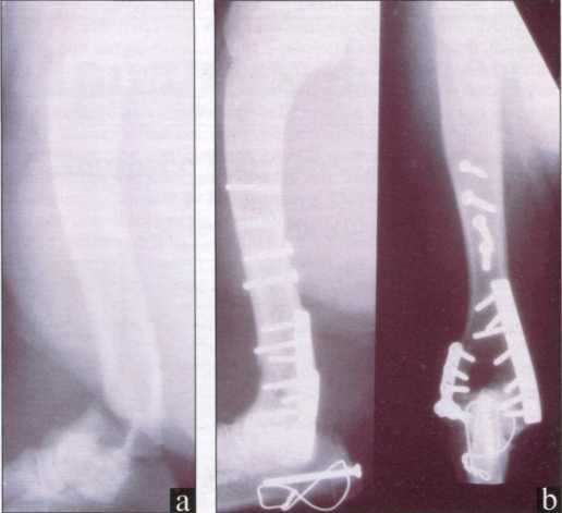
Figure 9 Mediolateral and craniocaudal radiographs of the humerus of a male Labrador Retriever cross after a road traffic injury.
(a) A supracondylar fracture is seen with several fissures in the proximal fragment.
(b) After operation. The fracture is fastened with a caudolaterally installed T-shaped bone plate, size 2,7 mm, installed by caudomedia! but the same size compressive bridge and 4 bone screws. An osteotomy of the olecranon was performed to gain access to the caudal distal part of the shadow bone. The olecranon is fastened with a bone screw with a diameter of 3.5 mm and a tightening wire bandage with a spoke.
After 10 weeks, the fracture healed.
Postoperative treatment
Immediately after the completion of the surgical fixation of the fracture, a control radiography is necessary. In cases of condylar or supracondylar fractures, the imposition of a soft bandage on a broken limb is indicated. The bandage is needed to prevent the development of limb edema and can be removed 3 days after the operation. For diaphyseal fractures or fractures in the proximal humerus, dressing after surgical fixation of the fracture is not required. All operated dogs are treated with painkillers for 1-5 days (as needed).
Physiotherapy is very important in the postoperative treatment of condylar fractures of the humerus. After such fractures in dogs, especially young dogs, there is often a limitation of mobility of the elbow joint as a result of primary or surgical tissue trauma, involvement of the condyle or epicondyle of the humerus in the fracture, and damage to the innervation of the limb. Even with good fracture bonding, the limited mobility of the elbow joint leads to a decrease in the weight load on the injured limb, which, in turn, further limits movement in the joint. Therefore, physiotherapy should be started as early as possible, since preventing the development of limited mobility is easier than restoring the lost freedom of movement in the joint.
Dog owners should be trained in basic physical therapy techniques. In practice, physiotherapy sessions should be carried out 2 times a day. These sessions should begin the next day after the surgical bonding of the fracture or immediately after the bandage is removed. Physiotherapy in the first 2 days after surgery involves the application of cold compresses to the injury site. Later, compresses can be made warm and supplemented with a gentle massage of the limb with fingers in the distal-proximal direction, especially in the area of injured and swollen tissues. At the same time, you should passively move the limb in the elbow, shoulder and carpal joints. After the dog has the opportunity to load the limb, physiotherapy can be replaced by walking on a leash. Walking up and down stairs, up and down steps, helps to restore mobility in the elbow joint especially well. such walks require more mobility in the elbow joint than when walking on a flat surface.
Restoration of joint mobility after surgical treatment in young dogs should be carried out within 2 weeks, and in adult dogs within 4-6 weeks. This ensures that the dog maintains good freedom of movement in the elbow joint.
Complications during treatment
In the treatment of fractures of the humerus, complications such as loss of mobility in the elbow joint, mixing of bone fragments in the fracture and its incorrect fusion, infections, iatrogenic denervation and, with fractures in the joints, degenerative diseases of the elbow or shoulder joints are possible. The frequency of complications after treatment of condylar fractures of the humerus is especially high in dogs with incomplete ossification of the condyle. This is probably due to sclerosis of the bone tissue in the condyle (Figure 3d). In case of poor fixation of the fracture, additional surgery may be required. Some dogs with incurable humerus fractures can regain use of the injured limb (albeit with lameness) within 4 to 12 weeks after surgery. If there is no function in the injured limb and its pain persists after surgical repair of the fracture or conservative treatment, arthrodesis of the elbow joint or amputation of this limb may be necessary.
LISTUSEDLITERATURE
- Johnson, J. A., Austin, C., Breur, G. J. Incidence of canine appendicular musculoskeletal disorders in 16 veterinary teaching hospitals from 1980 to 1989. Veterinary and Comparative Orthopaedics and Traumatology 1994; 7:56-69.
- Bardet, J. F., Hohn, R. B., Rudy, R. L., Olmstead, M. L. Fractures of the humerus in dogs and cats. A retrospective study of 130 cases. Veterinary Surgery 1983; 12:73-77.
- Matthiesen, D. T., Walter, M. Surgical management of distal humeral fractures. Compendium on Continuing Education for the Practicing Veterinarian 1984; 6:1027-36.
- Cockett P. A., Clayton Jones D. G. The incidence of humeral condylar fractures in the dog: a survey of seventy-nine cases. Journal of Small Animal Practice 1985; 26:437-44.
- Vannini, R., Olmstead, M. L., Smeak, D. D. An epidemiological study of 151 distal humeral fractures in dogs and cats. Journal of the American Animal Hospital Association 1988; 24:531-36.
- Vannini, R., Smeak,'D. D., Olmstead, M. L. Evaluation of surgical repair of 135 distal humeral fractures in dogs and cats. Journal of the American Animal Hospital Association 1988; 24:537-45.
- Ririnvik. A. M. Risk factors for humeral condylar fractures in the dog: a retrospective study. Journal of Small Animal Practice 1993; 34:277-82.
- Marcellin-Little, D. J., DeYoung, D. J., Ferris, K. K., Berry, S. M. Incomplete ossification of the humeral condyle in spaniels. Veterinary Surgery 1994; 23:475-87.
- Vannini, R., Olmstead, M. L., Smeak, D. D. Humeral condylar fractures caused by minor trauma in 20 adult dogs. Journal of the American Animal Hospital Association 1988; 24:355-62.
- Anderson, T. J., Carmichael S., Miller A. Intercondylar humeral fracture in the dog: a review of 20 cases. Journal of Small Animal Practice 1990;31:437-42.
- Kaderly, R. E., Lamothe M. Incomplete humeral condylar fracture due to minor trauma in a mature cocker spaniel. Journal of the American Animal Hospital Association 1992; 28:361-64.
- Denny, H. R. Condylar fractures of the humerus in the dog; a review of 133 cases. Journal of Small Animal Practice 1983; 24:185-97.
- Cook, J. L., Jordan, R. C. What is your diagnosis? Journal of the American Veterinary Medical Association 1997; 210:329-30.
- Brown, D. C., Conzemius, M. G., Shofer, F. S. Body weights as a predisposing factor for humeral condylar fractures, cranial cruciate rupture and intervertebral disc disease in cocker spaniels. Veterinary and Comparative Orthopedics and Traumatology 1996; 9:75-78.
- Marretta, S., Schrader, S. Physeal injuries in the dog: a review of 135 cases. Journal of the American Veterinary Medical Association 1983; 182:708-10.
- Marcellin-Little, D. J. Letter to the Editor. Another consideration for radiographic diagnosis. Journal of the American Veterinary Medical Association 1997; 210:1264.
- Pernell, R. T., Dunstan, R. W., DeCamp, C. E. Aneurysmal bone cyst in a six-month-old dog. Journal of the American Veterinary Medical Association 1992; 201: 1897-99.
- Dueland, R. Triceps tenotomy approach for distal fractures of the canine humerus. Journal of the American Veterinary Medical Association 1974; 165:82-86.
- Payne-Johnson, M., Lewis, D. G. A technique for fixation of intercondylar humeral fractures in immature small dogs. Journal of Small Animal Practice 1981; 22:293-299.
- Morshead, D., Stambaugh, J. E. Kirschner wire fixation of lateral humeral condylar fractures in small dogs. Veterinary Surgery 1984; 13:1-5.
- Klause, S. E., Schwarz, P. D., Egger, E. L., Piermattei, D. L. A modification of the unilateral type I external skeletal fixator configuration for primary or secondary support of supracondylar humeral and femoral fractures. Veterinary and Comparative Orthopedics and Traumatology 1990; 3:130-34.
- Aron, D. N., Foutz, T. L., Keller, W. G., Brown, J. Experimental and clinical experience with an 1M pin external skeletal fixator tie-in configuration. Veterinary and Comparative Orthopedics and Traumatology 1991; 4:86-94.
- Durall, I., Diaz, M. C., Morales, 1. Interlocking nail stabilization of humeral fractures. Initial experience in seven clinical cases. Veterinary and Comparative Orthopedics and Traumatology 1994; 7:3-8.
- Dueland, R. T. Interlocking nail fixation of humeral fractures. Proceedings of the 6th Annual Symposium of the American College of Veterinary Surgery 1996; 230.
For the treatment of fractures, the use of an immobilizing dressing (gypsum) has traditionally been used, this method of treatment has a number of disadvantages - the development of atrophy of the muscles of the limb, frequent malunion of bones, the formation of bedsores under the bandage, impaired blood supply to bones and soft tissues. All these complications led to the abandonment of the widespread use of gypsum for the treatment of fractures, so now this treatment method is used only for the treatment of cracks. A more modern method of treating fractures is osteosynthesis- surgery for surgical comparison of bone fragments with the use of fixing metal structures.
Types of osteosynthesis:
1. Intramedullary osteosynthesis - used to treat fractures of long bones. With this method, a special pin or needle is installed inside the bone. But there are limitations to this method - for example, it is not suitable for the treatment of fractures of the pelvis, skull, spine, jaw, as well as for the treatment of comminuted fractures.
ferret hip fracture

The use of intramedullary osteosynthesis for hip fracture
2. Bone osteosynthesis - with this method, a metal plate is attached to the bones with the help of special bolts. As a result, good stabilization of bone fragments is achieved. This method can treat not only fractures of tubular bones, but also injuries of the pelvis, skull, spine, scapula, etc. The negative side of this method is the rather high cost of the operation associated with the use of expensive materials (plates, bolts and special tools).

Fracture of the forearm in a dog

Bone osteosynthesis

Gunshot wound to the lower jaw with a fracture of both branches of the lower jaw

View after osteosynthesis
3. Extrafocal osteosynthesis - is used to treat not only fractures, but also dislocations, and consists in passing the spokes through the bone above and below the fracture site with their subsequent fixation from the outside with a special polymer. The advantages of this method are the relative cheapness of consumables, the speed of the operation, and the reliability of fixing debris. The disadvantage of this method is the impossibility of applying an external fixation device in large and giant breeds of dogs.

X-ray after extrafocal osteosynthesis
4. Combined osteosynthesis - consists in the use of several of the above methods and is used mainly for complex comminuted fractures.

A cat with a compound comminuted fracture of the femur

Cat after combined osteosynthesis

Intercondylar fracture of the humerus in a dog

After osteosynthesis
Separately, it is worth considering fractures of the pelvis. As a rule, such injuries are received by dogs as a result of car accidents, and cats by falling from a great height. In case of damage to the pelvic bones, fractures are usually multiple, which makes them the most difficult in the practice of a traumatologist.

Multiple pelvic fractures in a dog. On the right - a fracture of the pubic and ischial bones, on the left - a fracture of the acetabulum.

The same dog after osteosynthesis

Use of a compression plate for a complex fracture of the acetabulum
Our veterinary clinic has accumulated extensive experience in the use of all types of osteosynthesis in animals of all sizes, which allows us to approach the treatment of each case individually and recommend the most optimal method of reconstructive surgery.
Prices, rub.
The price does not include consumables and additional work
Question answerIs it possible to fix an old fracture (the radius of the front right paw in a dog)? If so, what is the name of this operation? A week later, we were booked in for an examination and an x-ray of an old fracture, we are waiting for what they say. But I would also like to get an answer to the question above ... The fracture has grown together crookedly, the dog is from the street. Julia
Q: Is it possible to fix an old fracture in a dog?
Hello! Maybe. This is metal osteosynthesis. But the only way to tell for sure is from the picture.
Hello. Tell me the approximate amount of total expenses, including additional ones, for prosthetics of the cat's paw. Amputated as a result of falling into a trap, in the area of the wrist.
Question: Can you tell me the approximate amount for prosthetics for a cat's paw?
Hello! For prosthetics please email us. [email protected] with a note to Sergey Sergeevich Gorshkov. It needs to be reviewed and reviewed. On the offhand, no one will say the approximate cost.
Anatomical and topographic data of the dog. Prevention of surgical infection, sterilization of instruments and materials. Preparation of the animal for surgery and its implementation by connecting bone fragments with plates. Possible complications and their elimination.
FSBEI HPE “St. Petersburg State Academy of Veterinary Medicine”
Department of Operative Surgery with the basics of topographic anatomy of animals
Course work
Operations on limbs
(osteosynthesis on the pelvic limb with a fracture of the femur in a dog)
Completed by: 3rd year student of 22 groups
Kantserova Anastasia Pavlovna
St. Petersburg 2012
1. Operation name
2. Goals of the operation
3. General information about the animal
8. Pain relief
9. Technique of the operation
11. Postoperative animal care
Conclusion
Bibliography
1. Operation name
Osteosynthesis (osteosynthesis; Greek osteon bone + synthesis connection) is the connection of bone fragments. There are two types of osteosynthesis - submersible osteosynthesis and external transosseous osteosynthesis. With submersible osteosynthesis, fixators connecting bone fragments are installed directly in the area of the fracture. External osteosynthesis is performed using various devices located above the skin and fixing bone fragments using pins and rods. The purpose of osteosynthesis is stable fixation of bone fragments in the correct position until their consolidation.
2. Goals of the operation
The purpose of osteosynthesis is to ensure stable fixation of fragments in the correct position while maintaining the functional axis of the segment, stabilization of the fracture zone until complete fusion. Basically, there are two types of treatment - surgical and conservative. The purpose of these types of treatment is to create conditions for restoring the integrity of damaged bone structures and surrounding tissues, as well as restoring the function of the damaged limb segment. With operational treatment methods fractures, traumatologists, as a rule, act directly on bone fragments. Conservative treatment is treatment without surgery, the doctor does not act on bone fragments, this effect occurs indirectly.
3. General information about the animal
Type, gender: dog, male.
Nickname: Bob
Color, markings: black
Breed: outbred
Age: about 4 years old
Height, weight: at the withers 65 cm, 30 kg
Preliminary diagnosis: fracture of the tibia of the right hind limb
Diagnosis at follow-up: fracture of 1/3 of the distal tibia of the right pelvic limb
Owner: homeless animal
There is no information about the conditions of keeping and feeding, as the animal was found on the street. Data on previous diseases and vaccinations are also unknown.
General study of the animal.
Determining the habitus of an animal:
Body position in space: forced, recumbent
Body type: average
Fatness: unsatisfactory
Temperament: phlegmatic
Constitution: rough
The pathological focus is located in the region of the right pelvic limb. There you can see a well-defined hyperemia, swelling, palpation of an increase in local temperature, numbness of the site, slight crepitus.
4. Fixation and location of the operation
Fixation is the strengthening of animals in a certain position in order to protect people conducting medical work from injuries from the patient, saving the life and health of the patient himself and preventing the destruction of surrounding structures by large and strong animals.
In dogs, so that they cannot bite, their mouths are tied with a strip of gauze, a gauze bandage or braid. Covering the mouth with a screw, its ends are first tied in the submandibular space with one simple knot, then the bandage is finally fixed on the back of the head with a marine knot.
Dogs are usually fixed on the table, giving them the necessary position. A simple operating table for small animals is made of wood: painted with white oil or enamel paint. The lid of the table should be concave inward or have a small recess in the middle with drains for liquid. Several holes are drilled in it for tying the mouth with straps (braid) used to fix dogs. Under the table, on its crossbars, a shelf is arranged in the middle, on which a basin is placed to drain liquids from the table top into it and collect the used dressing material.
Rice. 9. Strengthening the dog on the table: 1 - dorsal position; 2 - side; 3 - abdominal.
To strengthen the dog on the table in the dorsal position, a rope (braid) is tied or fixed with a rope loop to its thoracic limbs in the forearm area. The rope from each limb is passed between the limbs and chest and further under the back of the animal on the opposite side of the table to the corresponding hole; by pulling the rope, the limb of the dog is brought closer to the chest, after which the rope is tied. The pelvic limbs are extended and both are tied to the back of the table frame.
For the duration of the operation, cats are placed in special leather or dense fabric bags or wrapped in a piece of dense material, leaving the area necessary for the operation open. Even better, with any method of fixation, put on all the limbs of the cat special bags (stockings) made of durable fabric and then fix them accordingly.
During this operation, the animal was fixed in a lateral position.
5. Anatomical and topographic data
Musculature of the pelvic limb of a dog. A - from the lateral side B - from the medial side 1. sartorius muscle 2. semitendinosus muscle 3. biceps femoris muscle 4. cranial tibial muscle 5. long extensor of the fingers 6. long peroneal muscle 7. long flexor of the thumb 8. short flexor of the fingers 9 calf muscle 10. Achilles tendon 11. interosseous muscles 12. short extensor of fingers 13. short peroneal muscle 14. long flexor of fingers 15. slender muscle
The affected area is localized on the right pelvic limb. The fracture is on the tibia. It is surrounded by muscles:
Sartorius
Cranial tibialis muscle
Long finger extensor
Peroneus longus muscle
flexor thumb longus
Short finger flexor
Muscles are innervated by the tibial and peroneal nerves.
Vessels supplying muscles:
Posterior tibial artery
Anterior tibial artery
External iliac vein
Medial vein of saphenous
Caudal femoral vein
6. Instruments, dressings, medicines
Pointed scalpel, straight blunt and pointed scissors, wound hooks, anatomical and surgical tweezers, hemostatic forceps, needle holder, surgical needles - curved, semicircular, 5- and 10-gram syringes, injection needles, sterilizers for instruments and syringes, 0.5% novocaine solution, 5% alcohol solution of iodine, 0.5% ammonia solution, dressing material (sterile bandages, tampons, cotton wool), sterile PHA threads, polysorb, means for fixing animals, small surgical operating tables of the Vinogradov type, fixing elements: plates , screws, wrench for bending records, screwdriver for driving screws.
7. Prevention of surgical infection
STERILIZATION OF INSTRUMENTS
Basically, there are two ways to sterilize instruments: the action of high temperatures (boiling, sealing, etc.) and "cold" - in disinfectant solutions.
To sterilize instruments by boiling, simple or electric sterilizers are used (Fig. 22), which have a removable grill with handles. Sterilization is carried out in ordinary water with the addition of alkalis: 1% sodium carbonate; 3% sodium tetraborate (borax), 0.1% sodium hydroxide. The duration of boiling depends on the alkali dissolved in water: with sodium carbonate - 15 minutes, with borax - 20, with caustic soda - 10 minutes. Alkalis prevent metal corrosion, enhance sterilization efficiency and reduce boiling time.
The order of sterilization: the solution is brought to a boil, during this period the water is released from the oxygen dissolved in it and neutralized with alkali. Instruments are checked for suitability before sterilization. If they were covered with petroleum jelly, then it is wiped with alcohol or ether. The cutting part of the scalpel is pre-wrapped in gauze. Surgical needles are strung on a piece of gauze so that they are not "lost" in the sterilizer if there are a lot of instruments.
At the end of sterilization, the instruments are removed with the sterilizer grill and laid out on an instrument table covered in three rows with a sterile sheet or towel. At the same time, a certain order is observed - tools of the same type are placed in one place and in a certain sequence characteristic of each operation. The gauze in which the scalpels were wrapped should be unrolled. The laid out tools are covered with a sterile sheet or towel.
Used instruments (after opening abscesses, working with cadaveric material) are boiled (at least 30 minutes) in an alkaline liquid with the addition of 2% lysol or carbolic acid.
Glass objects (syringes, etc.) are placed in the sterilizer disassembled before it is heated. Syringes and glassware for anesthetic solutions are boiled in distilled water, since alkaline solutions contribute to the decomposition of some local anesthetics.
Sterilization of instruments by flombing (burning)
The disassembled tool is laid out in a clean enameled basin or bath, the required amount of alcohol is poured and it is lit. During the period of alcohol burning, it is advisable to turn the instrument over, since it cannot be sterilized well at the points of contact with the bottom. This method is used when providing emergency surgical care, as well as for sterilizing enameled dishes and tools that do not fit into the sterilizer due to their dimensions. Instruments are also sterilized in special cabinets at a temperature of 150-160 C for 20-30 minutes.
Sometimes in production conditions, tools are sterilized with antiseptic solutions. To do this, the tools are immersed for 30-40 minutes in one of the following solutions: 1% alcohol solution of brilliant green; ethacridine solution 1:500; 3--5% solution of carbolic acid; 1--2% Lysol solution or Karetnikov's liquid (formalin 20.0, carbolic acid 3.0, carbon dioxide 14.0, distilled water 1 l).
Rubber items are sterilized by boiling in distilled water. To do this, they are wrapped in gauze (so as not to burn) and boiled for 30 minutes or cold in formalin vapor.
Tool storage.
All instruments after the operation are thoroughly washed, sterilized and dried. Then they are laid out in a dry cabinet. To prevent rust on the instruments, a vessel half-filled with calcium chloride is placed in the cabinet. Injection needles can be stored in Nikiforov's liquid (alcohol and ether equally), while mandrin should be inserted into each needle. Dark spots or rust that has formed on tools can be removed with 2:1 chalk and ammonia. Rubber objects are placed separately from metal instruments. Do not store instruments together with iodine preparations, acids, etc.
Suture material and methods of its sterilization
Currently, the classification of suture material mainly takes into account two features: the ability to biodegrade and the structure of the thread.
According to the ability to biodegradation, there are:
absorbable materials (catgut, collagen, occelon, kacelon, vicryl, dexon, etc.);
non-absorbable materials (silk, nylon, lavsan, nylon, prolene, polyprolene, etc.).
According to the structure of the thread, they distinguish:
monofilament - is a homogeneous structure with a smooth surface;
shedding - in cross section it consists of many threads (twisted, braided, complex threads).
The following requirements are imposed on modern suture material:
Biocompatibility - the absence of toxic, allergenic, teratogenic effects of the suture thread on body tissues. Ideally, there should be no reaction to the suture material.
Biodegradation - the ability of the suture material to disintegrate and be excreted from the body. The suture should hold the tissue until a scar forms and then become unnecessary. At the same time, the rate of biodegradation should not exceed the rate of scar formation, in addition, the suture material should be atraumatic.
For the imposition of surgical sutures, linen and cotton threads are also used.
Silk sterilization
Silk threads are produced in bobbins (non-sterile) or in ampoules (sterile). Silk wound on glass coils or glass with polished edges is boiled in distilled water for 30-40 minutes. Store in 96° alcohol or in Nikiforov's liquid.
Sterilize silk and in solutions.
Sadovsky's method. Skeins of silk are placed for 15 minutes in a 0.5% solution of ammonia, and then for 15 minutes in a 2% solution of formalin in 70% alcohol.
Tour method. Silk is placed for 24-48 hours in a 1% alcohol solution of iodine. Store in the same solution.
Sterilization of cotton and linen threads.
These threads are less durable than silk threads. They are usually used to close skin defects in small animals, on the intestinal wall (ground floor), peritoneum. Use threads No. 10-20. They are sterilized according to the Sadovsky method or immersed for 24 hours in a 4% formalin solution.
Sterilization of catgut.
Catgut is made from the submucosal and partially muscular layers of the intestines of small cattle, and therefore requires special careful processing. Depending on the caliber, it is absorbed in the tissues of the animal body from 7 to 30 days.
Sterilization using high temperature is excluded. It is mainly used for submerged seams. It is produced in coils that require sterilization, or sterile - in sealed ampoules.
Gubarev's method. Loosely wound catgut on coils is degreased for 12-24 hours in ether or gasoline and sterilized in an alcoholic solution of iodine (1 g of iodine, 2 g of potassium iodide, 100 g of 95 ° ethyl alcohol) for 14 days, which is replaced with fresh in 7 days.
Method Ride. Without preliminary degreasing, the catgut is immersed in a 4% aqueous solution of formalin for 3 days.
Sadovsky-Kotylev method. The catgut is placed for 30 minutes in a 0.5% solution of ammonia, then transferred for 30 minutes to a 2% solution of formalin in 65 ° alcohol, in which it is stored until use.
Chubar's method. Catgut is immersed for 3 days in a liquid consisting of rectified alcohol 70 °, 200.0; glycerin - 5.0; tinctures of iodine - 8.0 and potassium iodide - 6.0. In this liquid, catgut is stored for a long time.
Sterilization of synthetic threads.
This material is sterilized by boiling in distilled water for 20 minutes. Metal wires and staples, as well as pins for connecting bones, are sterilized by boiling, usually together with instruments.
Sterilization of dressings, underwear and surgical items
Sterilization by autoclaving. Dressings (bandages, napkins, splints, compresses, tampons, etc.) and surgical underwear (gowns, sheets, towels, caps) are sterilized in autoclaves under pressure. Sometimes porcelain and glassware, enameled basins, solutions, etc. are placed there. Before autoclaving, the material and linen are loosely placed in biks (Fig. 23). Before placing the bixes in the autoclave, open the side holes, tightly close the lid. If there are no bixes, then surgical items are placed in canvas bags or bags. A pressure of 0.5 atm corresponds to a temperature of 115°C; 1 atm - 120; 2 atm - 134 ° C.
Before using the autoclave, close the release valve of the water-steam chamber, open the lid of the autoclave, pour water through the funnel to 2/3 of the level of the water-gauge glass, close the lid tightly and carefully tighten the bolts, after checking the tightness, turn on the heating source and release steam for 15-20 minutes; close the valve and raise the pressure to the level necessary for sterilization. Sterilization control is carried out by placing in the bix substances whose melting point is above 100 ° C.
After sterilization is completed, the autoclave is turned off, the drain valve is slowly opened, steam is gradually released, reducing the pressure, the autoclave lid is opened, the bixes are removed and the holes in them are immediately closed, the autoclave lid is closed.
Sterilization with flowing steam is carried out in a special Koch sterilizer, and in its absence, in a bucket or pan with a lid. They are filled with water to 1/3 of the height. The beginning of sterilization is considered from the moment steam is released, the temperature rises to 100 ° C, the duration is at least 30 minutes.
During sterilization by ironing, the temperature is brought to 100 ° C, the duration is at least 30 minutes.
During sterilization by ironing, the temperature is brought up to 150 ° C. Before sterilization, sheets, gauze, napkins are moistened with water and ironed at a speed of no more than 50 cm per minute, passing through the same place 2-3 times on both sides. The ironed material is folded with sterile tweezers and placed in a sterile bix or left wrapped in a sheet.
Preparing the animal and the surgeon for surgery
Preparing the animal for surgery.
For a favorable outcome of the operation, the preparation of the animal for it is important. Before the operation, the animal undergoes clinical studies, in particular, body temperature, respiration, and pulse rate are measured. It is impossible to perform the operation in animals with elevated temperature, it is also not recommended to perform it in the presence of infectious diseases, in malnourished animals. If the operation is not carried out urgently, then before it the animal is reduced to feed and, if possible, then a starvation diet is prescribed for no more than 12 hours.
When performing an operation under anesthesia, it should be borne in mind that some drugs, such as rometar, in the second half of pregnancy can cause fetal death. With a favorable outcome in these cases, surgery can be performed under local anesthesia, since it has been established that it does not affect the development of the fetus.
Before the operation, the animals are walked in order to free the large intestine, clean or partially anesthetize.
Operating field preparation
Preparation of the operation field is carried out in four stages: mechanical cleaning, degreasing, antiseptic treatment (asepticization), isolation of the operation field.
Mechanical cleaning includes washing with soap (preferably household soap), removing hair by shaving or clipping. In this case, the size of the prepared field should be sufficient to ensure sterile conditions for the operation. Mechanical cleaning is a particularly important stage in the preparation of the operation field and must be carried out with particular care, since it is due to it that the main amount of dirt and microorganisms is removed.
Preference is given to shaving, since asepticization with this method is more thorough. In practice, a safety razor is most often used. It has been established that hair shaving is best done on the eve of the operation, which allows not only to thoroughly remove the hair, but also to wash the surgical field well, which is usually heavily contaminated. In addition, skin irritation observed after shaving disappears by the time of surgery, as a result of which the skin becomes less sensitive to iodine solution and dermatitis develops less frequently. Accidental wounds of the skin during shaving by the time of the operation have time to become covered with a dense scab due to clotted blood.
Degreasing of the surgical field is carried out with a sterile gauze swab soaked in a 0.5% solution of ammonia or gasoline for 1-2 minutes. The fat-free operation field is treated with an antiseptic according to one of the following methods.
The Filonchikov-Grossich method, Its essence lies in the fact that the fat-free field is "tanned" and aseptic with a 5% iodine solution, first after mechanical cleaning, and then immediately before the incision or after infiltration anesthesia. In this case, the interval between treatments should be at least 5 minutes.
The Mouse method consists in the fact that after shaving, mechanical cleaning and degreasing, the operation field is treated with a 10% aqueous solution of potassium permanganate.
The Borchers method is based on the use of a 5% solution of formalin in 96% alcohol after mechanical cleaning, shaving and degreasing the skin. The method makes it possible to achieve (unlike most other methods) sterility in a protein medium (when contaminated with pus), since formalin retains its antiseptic properties.
Treatment of the surgical field with an antiseptic begins from the center (incision or puncture site) to the periphery. The exception is the presence of an open purulent focus, in which the treatment starts from the periphery and ends in the center.
field isolation operations are carried out using sterile sheets or oilcloths, which are attached to one another with special terminals (Backhaus) or pins.
Hand preparation before surgery.
During the operation, the surgeon's hands are in direct contact with the wound. It is known that the skin of the hands, like any other surface of the body, contains many microbes, most of which are pathogenic. Microbes find refuge in the excretory ducts of the sebaceous and sweat glands, in the subungual spaces, numerous furrows and skin folds. The skin of any part of the animal's body also contains a huge amount of them, so the preparation of the hands before the operation is of particular importance.
Hand treatment consists of three stages: a) mechanical cleaning; b) chemical disinfection; c) leather tanning. Some antiseptic substances often combine bactericidal and tanning properties (iodine alcohol solution, brilliant green solution, etc.), thus representing a bactericidal tanning agent or tanning antiseptic. Processing of hands is carried out from the fingertips and further to the elbows. For the mechanical processing of hands, it is necessary to have brushes made of plant material (agave leaves, palm trees, sabura), horsehair, synthetic, as well as soap, warm water, and basins.
Horsehair brushes do not tolerate boiling; they are treated with antiseptic substances. Brushes that were not in use are first washed thoroughly in warm water and soap, rinsed, and then immersed in a 3% solution of carbolic acid, a bactericide solution 1:3000 for 1 hour. They are also stored in these solutions.
When choosing one or another method of hand treatment, one should always keep in mind that hands cannot be absolutely sterile, they acquire only relative sterility for a certain time.
All methods of hand treatment are based on two principles: dehydration and skin tanning.
The chemicals used have bactericidal properties, affect the microbes that are on the surface of the skin, and tanning agents lead to the closure of the excretory ducts of the sweat and sebaceous glands and fix microorganisms in them.
The most accessible and simple to use are the following methods.
Alfeld method. After thorough mechanical cleaning in warm water with soap and a brush, hands are washed for 3 minutes. If hands are not wiped with a towel, then they are treated with 90 ° alcohol, if they are wiped with 70 ° alcohol. When the skin is dry, the subungual spaces are smeared with a 5% alcohol solution of iodine.
Olivkov's method consists in the fact that the hands are first washed for 5 minutes with hot water, soap and a brush, after which they are wiped with a towel and treated for 3 minutes with cotton wool soaked in a 1:3000 solution of iodine in alcohol.
In case of purulent operations, it is recommended to re-treat with iodized alcohol at a dilution of 1:1000.
Spasokukotsky-Kochergin method. According to this method, hands are washed with a 0.5% ammonia solution in two basins for 2.5 minutes or under a fluid stream of this solution. After the second wash, the liquid in the basin should remain clear. Otherwise, the washing is repeated and the hands are wiped with a towel. During the operation or when the hands are contaminated, the treatment is repeated.
The Napalkov method involves mechanical cleaning of hands with an aqueous solution of caustic potassium 1: 2000 with brushes for 5 minutes or in basins with napkins. Then the hands are wiped with a towel and treated with denatured alcohol for 3-5 minutes. Subungual spaces and skin folds are treated with 5% tincture of iodine.
Kiyashev's method is based on the use of the washing properties of a 0.5% ammonia solution, in which hands are washed with brushes for 5 minutes and wiped with a towel. Finished with a 3% zinc sulfate solution (3 min). Subungual spaces and nail beds are treated with a 5% iodine solution.
All of the above methods provide sterility of the skin of the hands for 20-30 minutes.
Currently, new bacteriostatic drugs are used that do not cause irritation and inflammation of the skin.
Zerigel. On clean, dry hands, apply 3–4 g of the drug and rub thoroughly for 8–10 s. Then dry hands for 2-3 minutes. If the film slips, re-treatment is not required. Sterility is ensured for 2 hours.
Also proposed is the treatment of hands with a 0.5% solution of catapol (sterility of the skin of the hands up to 3 hours) and a solution of chlorhexidine bigluconate in 70% alcohol at a dilution of 1:40 with an active substance concentration of 0.5%. Sterile hand skin remains for 4 hours.
Aerosols are also used to treat the skin of the hands: Septonex, etc.
Surgical gloves
None of the methods of processing hands brings them to a state of absolute sterility, therefore gloves are the only means by which sterility is ensured in the bacteriological sense of the word; This is especially necessary when carrying out operations for purulent-putrefactive processes, as well as when performing abdominal operations in small animals.
Since the integrity of the gloves cannot be guaranteed, it is necessary to pre-treat the hands using one of the above methods in order to prevent the transfer of "glove juice" consisting of sweat, exfoliating epithelium and bacteria to the wound. Sterilize gloves by boiling in distilled water for 30 minutes, as well as by autoclaving and in solutions: bactericide 0.1% - 15 minutes, chlorocide 2% - 30 minutes or in formalin vapor - 24 hours. After purulent operations, gloves are washed without removing them from the hands in a 2% solution of lysol.
8. Pain relief
During this operation, drugs for anesthesia were used:
Zoletil 50, 25% - 4 ml, intramuscularly administered throughout the operation;
Propofol 1% - 56 ml was administered throughout the operation, intravenously
Anesthesia
In this operation, both conduction anesthesia and infiltration anesthesia can be used.
With infiltration anesthesia, a 0.25-0.5% solution of novocaine is usually used, and much less often other drugs of this group in the amount of 10-15 ml at a time. During conduction, the same anesthetics are used, but at a higher concentration - 3.4 or 5%, and the amount of anesthetic depends on the thickness of the nerve, its depth, and the accuracy of the doctor's topographic orientation.
9. Technique of the operation.
Rice. 1. Oblique fracture; osteosynthesis by interfragmentary compression using screws and a neutralizing plate; scheme.
Training. The patient is tied in a lateral position and additionally fixed with a rope loop passed over the back and through the groin. The operated limb is placed on top and placed on a pillow. The incision site is shaved, the surgical field is treated with 5% iodine solution, the field is covered with sterile napkins
The course of action. The skin incision begins at the greater trochanter and is carried along the anterior edge of the femur to the knee joint. Superficial fascia, interfascial adipose tissue and deep fascia are dissected along the cranial edge of the biceps femoris muscle, the incision is made of the same length. After a wide opening of the edges of the wound, the intermuscular sheet of the broad fascia of the thigh is dissected, located caudolaterally on the femur, at the site of its attachment, then the lateral vastus muscle of the thigh is separated from the bone and retracted cranially with a retractor. If bleeding occurs as a result of damage to the muscular branches of the femoral artery and vein in the distal part of the incision, the vessels are ligated or coagulated.
A better view of the lateral surface of the femoral body can be achieved by retracting the biceps femoris and the lateral head of the quadriceps femoris with a retractor.
Rice. 2 The skin and superficial fascia are dissected and widely opened: A - femur; a - biceps femoris; b - lateral head of the quadriceps muscle, covered with fascia, b "- intermediate head of the quadriceps muscle; c - large and short adductors; a - square muscle of the thigh; 1 - sciatic nerve; 2 - muscle vessels
Access can be extended cranially by mobilization of the intermediate head of the quadriceps muscle and caudally by subperiosteal elevation of the adductors, if appropriate.
The femur is long, subject to strong flexion, and its reconstruction requires good stability.
Rice. 3. Access to the diaphysis of the femur
Osteosynthesis with a plate. A plate (neutralizing, tightening or supporting) is applied from the craniolateral side and fixed on each of the main fragments with at least three, and preferably four screws. Only in case of fractures at the point of transition to the metaphysis, it is enough to screw in two screws into a short fragment. In case of a multiple fracture with the formation of fragments that are not supplied with blood, separated from the periosteum and muscle attachment points, the optimal solution is to apply a plate on the medial side of the fracture (medial support). It is achieved by precise repositioning and correct bending of the plate. The remaining defects are filled with autogenous spongy substance.
In case of comminuted fractures with damage to soft tissues, the fracture zone is not left free according to the principle of biological osteosynthesis, but is connected indirectly, with the distraction of the main fragment, with a long plate fixed in the peripheral area, which achieves greater rigidity (support function!).
The plate extending to the knee joint must be adapted to the curvature of the femur. It should not be located in the lateral recess of the patella joint, it is fixed deep on the cranial edge of the distal fragment.
Wound closure. After placing the intermuscular sheet of the broad fascia of the thigh in place, the edges of the wound of the deep and superficial fascia are adapted in layers with an interrupted suture (absorbable material). With sufficient access to the hip and knee joint, further wound closure is carried out as described above. Suture the skin. Treat the seam with a 5% iodine solution.
10. Possible complications, their prevention and elimination
1. Fat embolism. In connection with the spread of fatty particles in the bloodstream, fatty embolism of the systemic circulation, pulmonary circulation and a mixed form may occur. A combination of fat embolism with traumatic shock is possible.
Warning - The treatment of fat embolism is complex. Its main directions are: - treatment and prevention of cardiovascular insufficiency (cardiac, hormonal, antihistamine, vitamin, vasodilating drugs); - treatment and prevention of respiratory failure (oxygen therapy, intubation or tracheostomy with artificial ventilation of the lungs in severe forms); -- correction of water-salt, protein metabolism, acid-base balance, prevention and treatment of acute renal failure: glucose-salt solutions and low molecular weight dextrans intravenously, albumin and protein blood substitutes, sodium bicarbonate, trisbufer, vasodilators, osmotic diuretics (lasix), inhibitors proteases (trasilol, contrykal), vagosympathetic blockades, exchange blood transfusions, in severe renal failure - hemodialysis; - careful patient care, toilet skin, oral cavity, tracheobronchial tree; -- prevention and treatment of infectious complications (antibiotics, sulfonamides, nonspecific and specific globulins, etc.); - in the first hours after the onset of complications, lipostabil or Essentiale is administered 1 drop per day. Lipostabil restores the physiological dissolution of demulsified neutral blood fat in the next few hours and improves the general condition of patients.
2. Anaerobic infection (gas gangrene). Anaerobic infection of wounds is observed very rarely, is one of the most severe complications of osteosynthesis, gives a high percentage of deaths and often forces surgeons to resort to amputation. Although this complication is extremely rare, doctors should be well aware of it. Anaerobic infection occurs with extensive damage to large muscle masses, mainly with injuries to the foot, lower leg, thigh and gluteal region. Factors contributing to its development are contamination of wounds with earth; violation of blood supply, prolonged pulling of the limb with a hemostatic tourniquet; general weakening of the body caused by fatigue, cooling, malnutrition.
Treatment for anaerobic infection consists of a complex of therapeutic methods used simultaneously, but in a known sequence: - if surgical treatment has not been performed, then it should be carried out radically, opening all pockets; - to release edematous muscles from compression, incisions should be made along the axis of the affected limb segment, and the incisions should penetrate to the muscles (“lamp” incisions); - If an anaerobic infection develops in a wound that has already undergone surgical debridement, a second radical debridement should be performed. Suturing after surgical treatment is contraindicated.
3. Osteomyelitis - purulent inflammation of all elements of the bone, accompanied by necrosis of part of it. The reason for the development of post-traumatic, postoperative osteomyelitis is a massive injury with the presence of necrotic tissues and microbial contamination (Staphylococcus aureus, hemolytic streptococcus, etc.).
Prevention of postoperative osteomyelitis: - prophylactic administration of antibiotics; - performing surgical interventions only in the absence of inflammation or skin necrosis; - strict observance of asepsis and antisepsis; - atraumatic operation; - meticulous hemostasis; - suturing the wound without tension, and if necessary - holding laxative incisions; - implementation of active drainage of the wound for 24 - 48 hours.
Conservative treatment of osteomyelitis: - after sowing pus on the flora and its sensitivity to antibiotics, targeted antibiotic therapy is performed (intramuscularly, intravenously, intraosseously, intraarterially); - The wound must be irrigated with antiseptic fluids. Surgical treatment of postoperative forms of osteomyelitis includes the following activities: - dissection and excision of the purulent focus, its good drainage; -- removal of metal structures and spokes; -- wide opening of the sequester box, removal of necrotic tissues, pathological granulations, sequesters; in the future, in order to fix fragments, preference should be given to transosseous osteosynthesis with devices. The closure of bone tissue defects is carried out with the help of muscle plasty.
4. Suppuration of wounds. Purulent infection of wounds is the most common complication of both internal and transosseous osteosynthesis. Clinical signs of it develop in most cases in the first 5 - 6 days after surgery. In some cases, purulent processes may also occur at a later date, when areas of late (secondary) necrosis are the substrate for suppuration.
Treatment consists in the following: - evacuation of the wound discharge and the creation of conditions for a constant outflow of pus; - with the development of infection in a sutured wound - removal of sutures and wide dilution of the edges of the wound; - loose tamponade of the wound cavity with gauze swabs moistened with antiseptic solutions, hypertonic sodium chloride solution; - in the presence of purulent leakage - its wide opening and good drainage and washing with antiseptic solutions; - if the purulent process is supported by the presence of necrotic tissues, repeated radical surgical treatment is indicated; a patient with a pronounced manifestation of a purulent infection should be on bed rest and receive a high-calorie diet rich in proteins and vitamins; - when anemia is detected - transfusion of small doses of fresh blood (250 ml) with a substitution and stimulating purpose; - the use of antibiotics, which should be targeted, i.e., only those to which microbes isolated from wounds are sensitive should be used; -- The limb must be well immobilized.
5. Violation of blood and lymph circulation. Often, especially in the treatment of fractures of the femur, a significant swelling of the limb develops. After giving the limb an elevated position, it decreases, but does not completely disappear. The cause of edema can be inflammation around the pins, thrombophlebitis, lymphostasis, as well as too forced distraction. In these cases, gradual distraction, a dosed load on the limb and therapeutic exercises should be carried out.
6. Secondary displacement of fragments.
Secondary displacement of fragments occurs due to non-compliance with the technique of applying the apparatus. In some cases, the displacement of fragments occurs when they are not sufficiently fixed (weak tension and fastening of the spokes, threaded rods, and rings), in others, as a result of errors made in the extrafocal osteosynthesis technique, when additional displacements are not eliminated, but, on the contrary, are created. efforts. Incorrectly fused fractures may occur in patients with incompletely aligned fragments or with unrepaired secondary displacement. Premature removal of the apparatus often leads to the development of angular deformities. This happens in those cases when the regenerate has a weak mechanical strength and its restructuring has not been completed.
7. Violations of asepsis and antiseptics during the operation could lead to the introduction of pathological microflora into the body of the animal, which would cause an inflammatory process or sepsis.
8. Rejection of the base plate.
Prevention is the observance of septic, asepsis during surgery, the selection of alternative methods of treatment.
11. Postoperative care
Restriction of mobility (interfering dressings, cage or room maintenance) until complete consolidation of the fracture or osteosynthesis, respectively. Also, to prevent licking or breaking the integrity of the wound, a collar must be worn on the dog. Be careful on stairs, falling off furniture. Keep warm and calm.
Immediately after the operation, put a dropper with stabilizol up to 150-200 ml, Amoxicillin 3 ml subcutaneously, a hemostatic drug - Dicinon 2 ml intravenously. Course - Rimadyl half a tablet 2 times a day, 5 days; Calcitriol 1 capsule per day for 3 weeks. Treat the seam with 5% iodine solution, powder, bandage.
Every day, treat the seam with o, o5% chlorhexidine solution or rometar, terramycin spray.
Make a biochemical blood test, X-ray, remove the stitches after 2 weeks.
Conclusion
The operation was successful. There were no complications during and after the operation. The outcome of the operation is recovery.
During the operation, the doctor and medical staff tried to ensure maximum sterility, the general condition of the animal was monitored, anesthesia was carefully dosed.
The method of osteosynthesis with plates was chosen based on the fact that this was the most optimal way to connect bone fragments. It provides maximum strength, as well as economic benefits.
Also, for a faster formation of callus, the drug Calcitriol in capsules was prescribed for a three-week course.
fracture dog surgical operation
Bibliography
1. Zelenevsky N.V. "Workshop on veterinary anatomy" volume 1. M .: NiK - 2007. - 852s.
2. Kalashnik I.A. "Workshop on general and private veterinary surgery". M .: Agropromizdat - 1988 - 303s.
3. Lebedev A.V., V.Ya. Lukyanovsky, B.S. Semenov "General veterinary surgery". M.: Kolos - 2000-448s.
4. Mozgov I.E. "Pharmacology" Moscow Agropromizdat 1985, 414p.
5. Petrakov K.A., P.T. Salenko, S.M. Paninsky Operative surgery with topographic anatomy of animals. Moscow "Kolos" 2001 - 423 p.
6. Semenov B.S. "Workshop on general and private surgery". M.: Kolos, 2000 - 448s.
7. Semenov B.S., A.V. Lebedev, A.N. Eliseev "Private veterinary surgery". M.: Kolos, 1997 - 496s.
8. Tkachenko S.S. "Portal about bone surgery"
9. Volmerhaus B., J. Frewein "Anatomy of a dog and a cat." "Aquarium" Moscow 2003 - 580 p.
10. Shebets H., V. Brass "Operative surgery for dogs and cats" "Aquarium" Moscow 2001-511p.
Similar Documents
Main indications for cystotomy. Surgery protocol. Anatomical and topographic data of the operated area. Preparing for animal surgery. Sterilization of instruments, stages of operation. Postoperative care and management of the animal.
test, added 04/28/2015
Definition of indications for ovariohysterectomy. Etiology, differential diagnosis, prevention and systematization of the disease. Preparing the dog for surgery Fixation and anesthesia of the animal. Instrumentation and its sterilization. Plan and technique of operation.
term paper, added 11/27/2014
Indications and contraindications for castration of boars. Preparing the animal for surgery, fixing it during it. Preparation of the surgeon's hands, instruments, suture and dressing material. Anatomical and topographic data of the operated area.
term paper, added 12/03/2011
Castration of females: the purpose of the operation. Ways to fix the animal. Location of the operation. Anatomical and topographic data. Instruments, dressings, medicines. Prevention of surgical infection, anesthesia. Operation technique.
term paper, added 12/06/2011
General and private preparation of the animal for surgery. Preparation of the surgeon's hands, instruments, suture and dressing materials. Anatomical and topographic data of the operated area, stages of the operation. Measures to prevent postoperative complications.
term paper, added 02/03/2012
General and private preparation of the animal for surgery. Preparation of the surgeon's hands, instruments and materials. Anatomical and topographic data of the operated area, animal fixation and anesthesia. Postoperative treatment, feeding, care and maintenance of the animal.
case history, added 12/23/2014
Scar puncture is an emergency operation. General preparation of the animal (cow) for the operation. Sterilization of instruments. Anatomical and topographic data of the operated area. Operational access. postoperative treatment. Feeding, care and maintenance of the animal.
term paper, added 12/08/2011
Anatomical and topographic data of the operated area. Clinical data of the cat. Preparation of instruments, animal, surgical field and hands for surgery. The use of infiltration anesthesia for pain relief. Surgical technique and postoperative care.
term paper, added 09/01/2012
Indications and contraindications for surgery, general preparation of the animal for its implementation: stages, principles and features. Preparation of the surgeon's hands, instruments, suture and dressing materials. Anatomical and topographic data of sheep. The principles of anesthesia.
case history, added 11/30/2011
Indications and contraindications for surgery in the abdominal wall of the horse. Preparing the animal for surgery. Preparation of the surgeon's hands, instruments, suture, dressing material and surgical underwear. Anatomical and topographic data of the operated area.
As with humans, fractures in dogs and cats are not uncommon. Treatment of fractures in animals is comparable in complexity to that in humans, and is often much more complex and comparable in complexity to pediatric traumatology.
There are a huge number of different types of fractures, and each type requires a different approach.
Types of fractures
A bone fracture can have a very complex name (diagnosis), such as "open comminuted fracture of the right condyle of the femur." This name is associated with a complex classification of fractures.
When broken, parts of the broken bone are called fragments.
If the fragments have not shifted relative to each other after the fracture, then this is a fracture without displacement. If they have shifted in any direction, then this is a fracture with a displacement.
A type of fracture without displacement - crack, microfracture of the bone. With such a fracture, the fragments are not only not displaced, but the fracture itself is not visible on a regular x-ray. Such a fracture is a time bomb, it can grow together, or it can finally break or, in the worst case, become a constant source of inflammation and pain. Computed tomography is required to diagnose these fractures.
If sharp fragments, after displacement, pierced the surrounding muscles and skin and came out, then this open fracture, if the skin is not damaged, then such a fracture closed.
If the fragments broke obliquely, then the fracture oblique, If the fragments broke at an angle of 90 degrees, then straight(the easiest option), if the fragments broke in a spiral, then spiral fracture.
If small fragments are formed during the fracture, then such a fracture is called splintered.
The middle of a bone is called the diaphysis, and the end of the bone that meets another bone at a joint is called the epiphysis. If the bone is broken in the middle, then it is a fracture. diaphyseal. If the bone is broken at either end, then such a fracture is called epiphyseal.
Type of epiphyseal fracture articular fracture. With such a fracture, the epiphysis is broken inside the joint, and not only the bone and surrounding muscles are damaged, but also the joint, which significantly worsens the condition of the dog and can result in permanent lameness and arthrosis of the joint.
There are many types of articular fracture.
Detachments. There are separations of muscles from the bone or breaks of pieces of bone to which the muscle is attached.
Condylar fractures in which half of the articular surface (condyle) breaks off.
In complex joints, which are formed by several bones, one bone can break and the other is not damaged. This is incomplete fracture.
If there is only one fracture of the bone, then this simple fracture. If there are several fractures of one bone or the fracture is inside the joint, then such a fracture complicated. Difficult because the treatment of such a fracture requires more experience and knowledge of the doctor, and the operation is long in time.
After reading these classifications (which are far from all listed), one might think that anything and in any way can break down in an animal in accordance with the classification.
In real life, things are a little different. There are statistics and according to her data, animals break only a few bones in 70% of cases, the remaining 30% are rare.
Most often, cats and dogs break their paws, then the spine, and finally the bones of the skull.
If you take paws, then large bones and major joints break first of all.
If it's front paws, then it's
- fractures of the radius and ulna
- fractures of the radius and ulna and elbow joint
- humerus fractures
- fractures of the humerus and shoulder joint
even more rarely fractures in the wrist and metacarpus
If it's the hind legs, then
- femur fractures
- knee fractures
- fractures of the knee and femur
- fractures of the knee and tibia
- tibial fractures
- hip fractures
- pelvic fractures
- pelvic and hip fractures
even less often fractures in the area of the tarsus and metatarsus
In terms of fractures in the head area, the lower jaw leads.
In fractures of the spine, most often break
- first cervical vertebrae
- lumbar spine
- sacral spine
- thoracic spine
Working constantly with the same type of cases, the orthopedic veterinarian studies specific types of fractures in relation to certain bones or joints, studies specific methods for treating such fractures, and has a huge advantage over the general surgeon in the treatment of fractures in animals.
For a veterinary orthopedist, classifications and methods of treatment in relation to a specific joint, and not to a generalized concept of a bone or joint, come to the fore.


Incomplete fracture of a long tubular bone (greenstick)


Treatment of fractures in dogs and cats
The animal is examined by a doctor, if the cat or dog is in a non-life threatening condition, the fracture is diagnosed (X-rays and, in some cases, computed tomography) and the fracture is treated ( osteosynthesis).
Osteosynthesis - (ancient Greek ὀστέον - bone; σύνθεσις - connection) is a surgical operation to connect bone fragments using various fixing structures that ensure stable fixation of fragments in the correct position.
If there is a threat to the life of the patient, then such a patient is first “stabilized”, no matter how much time it takes, and then diagnostics of fractures and osteosynthesis are carried out.
Anesthesia plays an important role in the treatment of animals with fractures, especially severely injured animals. Without anesthesia, the animal may die from shock or simply suffer from severe pain.
The choice of the osteosynthesis method plays a very important role. The rate of fracture healing and recovery of the animal depends on how correctly the method is chosen.
When choosing a method, the materials used during the operation are also important. For example, a bone can be connected with a plate, but the plates, due to the large surface of contact with the bone, slow down the formation of callus. The imposition of the plate is a factor in slowing down the union. But there are plates made of very strong materials, which are smaller than conventional ones and have limited contact with the bone due to a specially designed rolling profile. Such plates do not affect the healing rate.
When performing osteosynthesis, the main task is to match the fragments as anatomically correctly as possible and fix them in this position 100% immobile. This is the only way to achieve the fastest healing of the fracture.
The second task facing the orthopedic surgeon is to perform the operation quickly and minimally traumatically. The speed is ensured by experience and clear planning of the course of the operation, as well as the chosen method of osteosynthesis.
It is important, during the operation, not to damage the nerves and blood vessels in the surrounding tissues. Their damage can lead to the loss of the paw.
A person is often put in a cast, an animal never. This is an unshakable rule that is constantly violated by general surgeons.
Plastering a dog or cat results in:
A. GYPSUM - MOBILITY OF FRAGMENTS - FALSE JOINT - TREATMENT FOR YEARS - RADICAL RECOVERY SURGERY - FREQUENTLY EUTHANASIA
The imposition of plaster on a dog or cat leads to the mobility of the fracture, its long-term non-union and the formation of a false joint. A plaster cast cannot tightly fix the short small and often crooked (unlike human) bones of cats and dogs. Dogs and cats cannot lie down and wait for the fusion, they want to run and loosen the already unreliable plaster cast. The bones at the fracture site are constantly moving and do not allow the formation of callus (fracture union). If the bones are constantly moving at the fracture site, then they “grind” to each other, cartilage is formed at their ends and a false joint occurs. Such a fracture may not grow together for years.
B. GYPSUM - NECROSIS (DEATHING) OF THE PAWS TISSUES - REMOVAL OF THE PAWS - OFTEN EUTANASIUM
Casting a dog or cat results in necrosis (death of the paw) and loss of the paw (removal).
Or to inflammation of the paw, suppuration, long-term treatment and removal of the paw in extreme cases.
People often euthanize animals because they are not ready to take care of a disabled animal.
C. GYPSUM - SEVERE DERMATITIS UNDER GYPSUM - SUPPRESSION - SKIN PLASTY - RADICAL RECOVERY SURGERY - FREQUENTLY LOSS OF PAWS AND EUTHANASIS
Casting is always accompanied by severe dermatitis of the skin under the cast (wool, moisture and dirt cannot simply be preserved under a cast, they destroy the skin and paw under the cast). This is followed by a long recovery of the skin, plastic, antibiotic treatment and a complex reconstructive operation.
People often despair and euthanize animals, because they are not ready to pay for a complex operation to restore a paw, they are not ready to take care of a cripple animal.
THUS, GYPSUM IN 99% OF CASES LEADS TO COMPLICATIONS AND RECOVERY SURGERY WITH A LOWER PROBABILITY OF COMPLETE RECOVERY OF THE PAWS.
60% of the work of orthopedists, plastic surgeons in our clinic is made up of alterations and retreatment after unsuccessful osteosynthesis and plastering at home and in conditions close to those of military field surgery (but these dogs were not on the battlefield), by doctors who do not have experience and knowledge to carry out such operations and do not have the necessary tools and consumables.
Features in the treatment of spinal fractures
The only difference is time.
The spinal cord and nerves can be destroyed irreversibly and the animal will remain disabled.
Time is not playing into your hands.
If the spine is damaged, there is a possibility of damage to the spinal cord by bone fragments and displaced vertebral bodies. The sooner the load on the spinal cord is removed, the faster the spinal cord will begin to recover and the less chance that irreversible damage to the spinal cord will occur.
Care after surgery
Care for a recovering pet depends on the type of fracture and the type of surgery that has been performed. Although most pets can be allowed to do some exercise almost immediately after surgery, it is important that unrestricted activity such as running or jumping is avoided. Sometimes it is required to keep a pet in a limited area, for example, in a small room. Physiotherapy and hydrotherapy may be recommended as part of a recovery program. In most cases, we re-examine the pet six to eight weeks after surgery. When x-rays show that the fracture has healed, normal activity can be resumed.
Risks and difficulties associated with the treatment of fractures
Even an operation performed flawlessly can carry the risk of certain complications such as infection and difficulty in bone healing. However, if the operation is performed by an experienced specialist, such complications are rare, and most pets recover without complications. Joint fractures can lead to osteoarthritis, which may require long-term follow-up, but most pets do not experience these complications. Before any treatment is started, all aspects of your pet's postoperative care, including the risk of complications, will be detailed during your initial consultation with the orthopedist.
prospects
Most pets gain full use of their limbs and can enjoy a normal life.
Why should you contact us for the treatment of a fracture in your pet?
- We have extensive experience in the successful treatment of various fractures;
- We are attentive to each patient;
- We use modern technologies and methods of treatment;
- Our experts regularly take part in veterinary conferences;