There are many questions about the structure of the eye. This organ is in second place after the brain in terms of the complexity of the structure in the human body. It is surprising that such a small organ of vision has a huge number of working systems and is distinguished by great functionality. The structure of the organ of vision implies the presence of more than two and a half million components, while in a short moment of time a large amount of information is processed. Due to the fact that the structure of the human eye involves coordinated work, and functions are performed. This is the key to clear vision.
The anatomy textbook diagram will tell you in detail about the structure of the human eye. There are several departments, each of which has its own functions:
- eyelids;
- eyelashes;
- sclera of the eye;
- cornea;
- limbo.
This is a small part of those departments that represent the human eye. The eye itself refers to the eyeball. It is presented in a spherical shape with irregular outlines. On average, the size is more than two tens of mm in an adult. The eyes are located in a special compartment of the bone type - the eye sockets. From the outside, the organ of vision is protected by eyelids, along the edges by special muscles that are responsible for the movement of the eyes and fiber related to the fatty type. On the reverse side is the central nerve, which supplies data to the brain.
Features of human vision are in the device of the process by which the image is formed. Initially, light passes through the cornea, which lines the outside of the eyeball. It focuses on the first level. Part of the iris scatters the rays, the rest passes through the pupil. Due to its adaptability, people can perceive objects in different lighting conditions.
The final refraction of the light beam occurs with the help of a lens. After that, the passage through the body of the vitreous type is carried out. The rays are scattered over the eye retina, which acts as a recipient, which converts the information received from the light stream into a nerve-type impulse. The image itself is formed due to the decoding of this impulse by the brain.
Eyelid Features
The external structure of the eye is associated with the formation of the eyelids. By them are meant special partitions. The main function is to protect the eyeball from external factors and injuries. For the most part, the eyelid is represented by muscle tissue. On the outside, it is lined with thin skin. Due to the fact that the tissues here are muscular, both eyelids have the possibility of free movement.
Due to the constant closing of the eyelids around the eyeball, moisturizing and removal of particles of a different origin occur. Within the science of the eye - ophthalmology, it is emphasized that the eyelids are an important element. The device of the eyes is made in such a way that any damage to the eyelids can provoke diseases.

In order to preserve the shape of the eyelid and make it durable, cartilage is “designed” by nature. This is a dense formation of collagen. Inside the cartilage are the meibomian glands, which produce a fat-based secret. It is required by the eyelids for a tighter closure.
From the inside, the conjunctiva of the eye is attached to the cartilage. The structure of the human eye suggests the presence of a special mucous membrane, which produces fluid. Without it, hydration would not be possible. This fluid helps the eyelid glide better over the surface of the eyeball. The vessels lining the eye are represented in the eyelid by a system with a large number of branches. Secular functions are controlled by three types of nerves.
Muscles of the eye
An important role that determines the structure and functions of the eye is assigned to the muscular body. It depends on them what position the eyeball will have, how it will function. Dozens of muscles are fixed on the outer and inner sides of the eyelids. However, most of the tasks are assigned to the muscular processes of the oblique and direct type.
Muscle groups come out of the tendon ring, which is hidden in the orbital depths. Above the muscle of the direct type, located on top, a muscle is also attached to the ring, the main function of which is the lifting of the eyelid, located on top.
The rectus muscles line the orbital walls, which surround the nerve from different sides. At the end of the muscles are shortened tendons. The structure of the sclera involves attaching them to the tissues. The rectus muscles at the same time help the eye to turn in a given direction.

The oblique muscle located below, which is still formed on the upper jaw, differs in its structure. This muscle has an upper direction in an oblique design and is located in the back. According to the science of the eyes, due to the consistency in the complex work of the muscles of the eye, the apple itself turns in the direction that the user requires. In addition, the work of two eyes at the same time is coordinated.
The structure and functions of the organ of vision suggest different types of membranes. Each performs its own functionality. It is not only about protection from factors of external origin, but also about coordinated work.
With the help of the fibrous membrane, the eye is protected from factors that can damage it from the outside. Actually, the choroid of the eye collects excess light rays, preventing them from reaching the retina, which lines the organ of vision, in full. The vascular membrane of the eye is also responsible for the distribution of the blood supply that the eyeball needs on different layers.
Another shell affects the depths of the eyes. It refers to the retina. This visual department has two pigment parts, which are located outside and inside. Inside, the retina also has two parts. One of them is equipped with elements that react to light, the other is deprived of them.
Small departments
The sclera is an important part of the visual organ. The sclera is the membrane that covers the eyeball almost completely (80 percent). Further, the sclera flows into the cornea from the front side. In the common people, the sclera is called the white of the eye. In this case, the sclera has a venous sinus in a circular design in a place where the anatomy suggests a connection with the cornea.
The cornea can be considered an extension of the sclera of the eyes. This element of the eyeball can be perceived as a plate that is colorless. The anterior corneal part is convex, and behind there is some depression. The edge is in contact with the body of the sclera. Some compare it to watch glass. Physics would classify the cornea as a lens, without which the visual process is impossible.
The next important physical department is the iris. It refers to the visible part of the choroid. It has a disc shape, in the center of which is the pupil, which is a hole. The iris determines the color of a person's eyes. It depends on how dense the stroma is and how much pigment is used in it.

When pigments are used a little and with fabrics of high friability, the iris often has a blue tint. If there is enough pigment, but the friability of the fabric is the same, a green tint may appear. Dense tissues with a small amount of pigment are characteristic of gray eyes. High density, coupled with a large amount of pigment, is found in owners of brown eyes.
The iris is not that thick. This is 0.2-0.4 tenths of a millimeter. On the surface in the anterior part there is a ciliary and pupillary girdle. A small circle of arteries is used to separate them. It is woven from the arteries of a thin size.
The ciliary body also has many elements. The ciliary body is located behind the iris. The main task of this part of the eye is the production of a special composition. By and large, the ciliary body is responsible for nourishing and filling with liquid the eye sections located in the anterior part. It is completely penetrated by the vessels of the eye. At the same time, the fluid that the ciliary body produces has a number of features.
In addition to a huge number of vessels, the ciliary body is distinguished by a developed muscular complex. Due to relaxation and contraction, the shape of the lens changes. With contraction, the lens increases in thickness, which means that the optical effect is enhanced. This is important for obtaining a high-quality image of objects that are located next to a person. If the muscles are relaxed, then the lens contracts in its thickness, and a person can distinguish objects located in the distance.
Additional parts
Under the concept of the lens, anatomy understands the body of a transparent color, which is located opposite the pupil. The lens is hidden in the depths of the eyeball. By and large, the lens can be considered a biological lens, which is distinguished by its double convex shape. It is the lens that plays the main role. Without its normal functioning, human vision will not be able to work properly. The lens is surrounded by the vitreous body and the iris. If a person does not suffer from developmental disorders, then the thickness of the lens in its maximum value can vary from three to five millimeters.
Another important section is the retina, which lines the inside of the eye. With its help, the projection of the existing image and its final processing are performed. In case of malfunctions, it can be pulled together by the epiretinal membrane. The epiretinal membrane is scar tissue that leads to the formation of folds and wrinkles. It is worth noting that the epiretinal membrane is often formed as a complication of some kind of eye disease. Most often, the epiretinal membrane is recorded in older people, starting from 65 years. At the same time, the epiretinal membrane has no sex dependence, and occurs equally often in men and women.

With the help of the retina, various information flows are formed into a common one. Here there are several stages of filtering and processing information by other departments that are present in the eyeball. As a result, an impulse is formed, which reaches the brain through the nerve endings.
The base of the retina is formed by two cell types. Cones and rods are photoreceptors and act as converters of light energy into "electricity". With a small number of light sources, rods are an important part of vision, and cones are mostly activated when there is sufficient light. Thanks to them, colors and fine details of objects are distinguished. The disadvantage of the retina is its loose fit to the sheath of blood vessels. As a result, detachment occurs with microtrauma, which causes eye diseases.
How light is changed and processed
The structure of light refraction in the human eye has a lens system. The first lens is the cornea of the eye. Due to this part, a person can see 190 degrees around him. With violations in the cornea, tunnel vision pathologies are formed. Finally, the beam of light is refracted by the eye lens, which is responsible for focusing the rays on a small area of the retina. The lens varies visual acuity, with changes comes nearsightedness or farsightedness.
With the help of accommodation structures, the intensity of the light that enters and focuses is regulated. The structure of the accommodation structure includes the iris, pupil, muscles of various types.
It is sometimes referred to as a lens. By changing the curvature, the human eye focuses on objects located near or far. The ciliary muscles are responsible for changing the curvature. The luminous flux is regulated due to changes in the pupillary diameter, which leads to the expansion or narrowing of the iris. Each of these processes is responsible for its own group of iris muscles.
The structure of the receptor type is represented by the retina, in which photoreceptor cells and neurons that approach them are located. The retina has a complex anatomical structure, it is heterogeneous. It has a blind spot and an area with increased sensitivity. It has ten layers. The main function of light information processing is assigned to photoreceptor cells, which have a rod and cone appearance.
The organ of vision is the most important of all human senses, because about 90% of information about the outside world a person receives through a visual analyzer or visual system.
The organ of vision is the most important of all human senses, because about 90% of information about the outside world a person receives through a visual analyzer or visual system. The main functions of the organ of vision are central, peripheral, color and binocular vision, as well as light perception.
A person sees not with his eyes, but through his eyes, from where information is transmitted through the optic nerve to certain areas of the occipital lobes of the cerebral cortex, where the picture of the outside world that we see is formed.
The structure of the visual system
The visual system consists of:
* Eyeball;
* Protective and auxiliary apparatus of the eyeball (eyelids, conjunctiva, lacrimal apparatus, oculomotor muscles and orbital fascia);
* Life support systems of the organ of vision (blood supply, production of intraocular fluid, regulation of hydro and hemodynamics);
* Conducting pathways - optic nerve, optic chiasm and optic tract;
* Occipital lobes of the cerebral cortex.
Eyeball
The eye has the shape of a sphere, so the allegory of an apple began to be applied to it. The eyeball is a very delicate structure, therefore it is located in the bony recess of the skull - the eye socket, where it is partially sheltered from possible damage.
The human eye is not quite the correct spherical shape. In newborns, its dimensions are (on average) along the sagittal axis 1.7 cm, in adults 2.5 cm. The mass of the eyeball of a newborn is up to 3 g, an adult - up to 7-8 g.
Features of the structure of the eyes in children
In newborns, the eyeball is relatively large, but short. By 7-8 years, the final size of the eyes is established. The newborn has a relatively larger and flatter cornea than adults. At birth, the shape of the lens is spherical; throughout life, it grows and becomes flatter. In newborns, there is little or no pigment in the stroma of the iris. The bluish color of the eyes is due to the translucent posterior pigment epithelium. When the pigment begins to appear in the iris, it takes on its own color.
The structure of the eyeball
The eye is located in the orbit and is surrounded by soft tissues (fatty tissue, muscles, nerves, etc.). In front, it is covered with conjunctiva and covered with eyelids.
Eyeball consists of three membranes (outer, middle and inner) and contents (vitreous body, lens, and aqueous humor of the anterior and posterior chambers of the eye).

Outer, or fibrous, shell of the eye represented by dense connective tissue. It consists of a transparent cornea in the anterior part of the eye and a white opaque sclera. With elastic properties, these two shells form the characteristic shape of the eye.
The function of the fibrous membrane is to conduct and refract light rays, as well as protect the contents of the eyeball from adverse external influences.
Cornea- transparent part (1/5) of the fibrous membrane. The transparency of the cornea is due to the uniqueness of its structure, in it all the cells are located in a strict optical order and there are no blood vessels in it.
The cornea is rich in nerve endings, so it is very sensitive. The impact of unfavorable external factors on the cornea causes a reflex contraction of the eyelids, providing protection for the eyeball. The cornea not only transmits, but also refracts light rays, it has a large refractive power.
Sclera- the opaque part of the fibrous membrane, which has a white color. Its thickness reaches 1 mm, and the thinnest part of the sclera is located at the exit of the optic nerve. The sclera consists mainly of dense fibers that give it strength. Six oculomotor muscles are attached to the sclera.
Functions of the sclera- protective and shaping. Numerous nerves and vessels pass through the sclera.
choroid, the middle layer, contains the blood vessels that carry blood to feed the eye. Just below the cornea, the choroid passes into the iris, which determines the color of the eyes. At its center is pupil. The function of this shell is to limit the entry of light into the eye at high brightness. This is achieved by constricting the pupil in high light and dilating in low light.
Behind the iris is located lens, similar to a biconvex lens that catches light as it passes through the pupil and focuses it on the retina. Around the lens, the choroid forms a ciliary body, in which the ciliary (ciliary) muscle is embedded, which regulates the curvature of the lens, which provides a clear and distinct vision of objects at different distances.

When this muscle is relaxed, the ciliary band attached to the ciliary body is stretched and the lens is flattened. Its curvature, and hence the refractive power, is minimal. In this state, the eye sees distant objects well.
In order to see near objects, the ciliary muscle contracts and the tension of the ciliary girdle weakens, so that the lens becomes more convex, hence more refractive.
This property of the lens to change its refractive power of the beam is called accommodation.

Inner shell eyes presented retina– highly differentiated nervous tissue. The retina of the eye is the front edge of the brain, an extremely complex formation both in structure and in function.
Interestingly, during embryonic development, the retina is formed from the same group of cells as the brain and spinal cord, so it is true to say that the retinal surface is an extension of the brain.
In the retina, light is converted into nerve impulses, which are transmitted along the nerve fibers to the brain. There they are analyzed, and the person perceives the image.
The main layer of the retina is a thin layer of light-sensitive cells - photoreceptors. They are of two types: responding to weak light (rods) and strong (cones).
Sticks there are about 130 million, and they are located throughout the retina, except for the very center. Thanks to them, a person sees objects on the periphery of the field of view, including in low light.
There are about 7 million cones. They are located mainly in the central zone of the retina, in the so-called yellow spot. The retina here is maximally thinned, all layers are missing, except for the layer of cones. A person sees best with a yellow spot: all light information that falls on this area of \u200b\u200bthe retina is transmitted most fully and without distortion. Only day and color vision is possible in this area.
Under the influence of light rays in photoreceptors, a photochemical reaction occurs (disintegration of visual pigments), as a result of which energy (electric potential) is released that carries visual information. This energy in the form of nervous excitation is transmitted to other layers of the retina - to bipolar cells, and then to ganglion cells. At the same time, due to the complex connections of these cells, random “noise” in the image is removed, weak contrasts are enhanced, moving objects are perceived more sharply.
Ultimately, all visual information in an encoded form is transmitted in the form of impulses along the fibers of the optic nerve to the brain, its highest instance - the posterior cortex, where the visual image is formed.

Interestingly, the rays of light, passing through the lens, are refracted and turned over, due to which an inverted reduced image of the object appears on the retina. Also, the picture from the retina of each eye enters the brain not entirely, but as if cut in half. However, we see the world normally.
Therefore, it is not so much in the eyes as in the brain. In essence, the eye is simply a perceiving and transmitting instrument. The brain cells, having received an inverted image, turn it over again, creating a true picture of the surrounding world.
Contents of the eyeball
The contents of the eyeball are the vitreous body, the lens, and the aqueous humor of the anterior and posterior chambers of the eye.
The vitreous body by weight and volume is approximately 2/3 of the eyeball and more than 99% consists of water, in which a small amount of protein, hyaluronic acid and electrolytes are dissolved. This is a transparent, avascular gelatinous formation that fills the space inside the eye.
The vitreous body is quite firmly connected with the ciliary body, the lens capsule, as well as with the retina near the dentate line and in the region of the optic nerve head. With age, the connection with the lens capsule weakens.
Auxiliary apparatus of the eye
The auxiliary apparatus of the eye includes the oculomotor muscles, lacrimal organs, as well as the eyelids and conjunctiva.

oculomotor muscles
The oculomotor muscles provide the mobility of the eyeball. There are six of them: four straight and two oblique.
The rectus muscles (superior, inferior, external, and internal) originate from a ring of tendons located at the apex of the orbit around the optic nerve and insert into the sclera.
The superior oblique muscle starts from the periosteum of the orbit above and medially from the visual opening, and, going somewhat backwards and downwards, is attached to the sclera.
The inferior oblique muscle originates from the medial wall of the orbit behind the inferior orbital fissure and inserts on the sclera.
The blood supply to the oculomotor muscles is carried out by the muscular branches of the ophthalmic artery.
The presence of two eyes allows us to make our vision stereoscopic (that is, to form a three-dimensional image).
Precise and well-coordinated work of the eye muscles allows us to see the world around us with two eyes, i.e. binocularly. In case of dysfunction of the muscles (for example, with paresis or paralysis of one of them), double vision occurs or the visual function of one of the eyes is suppressed.
It is also believed that the oculomotor muscles are involved in the process of adjusting the eye to the process of vision (accommodation). They compress or stretch the eyeball so that the rays coming from the observed objects, whether far or near, can hit the retina exactly. In this case, the lens provides finer adjustment.
Blood supply to the eye
The brain tissue that conducts nerve impulses from the retina to the visual cortex, as well as the visual cortex, normally almost everywhere has a good supply of arterial blood. Several large arteries that are part of the carotid and vertebrobasilar vascular systems participate in the blood supply of these brain structures.

Arterial blood supply to the brain and visual analyzer is carried out from three main sources - the right and left internal and external carotid arteries and the unpaired basilar artery. The latter is formed as a result of the fusion of the right and left vertebral arteries located in the transverse processes of the cervical vertebrae.
Almost the entire visual cortex and partly the cortex of the parietal and temporal lobes adjacent to it, as well as the occipital, midbrain and pontine oculomotor centers are supplied with blood due to the vertebrobasilar basin (vertebra - translated from Latin - vertebra).
In this regard, circulatory disorders in the vertebrobasilar system can cause dysfunction of both the visual and oculomotor systems.

Vertebrobasilar insufficiency, or vertebral artery syndrome, is a condition in which blood flow in the vertebral and basilar arteries is reduced. The cause of these disorders may be compression, increased tone of the vertebral artery, incl. as a result of compression by bone tissue (osteophytes, herniated disc, subluxation of the cervical vertebrae, etc.).
As you can see, our eyes are an exceptionally complex and amazing gift of nature. When all departments of the visual analyzer work harmoniously and without interference, we see the world around us clearly.
Treat your eyes carefully and carefully!
Undoubtedly, each of the sense organs is important and necessary for a person to fully perceive the world around him.
Vision allows people to see the world as it is - bright, diverse, unique.
Organ - vision
In the human organ - vision - one can distinguish the following components:
- The peripheral zone is responsible for the correct perception of the initial data. In turn, it is divided into:
- eyeball;
- protection system;
- accessory system;
- propulsion system.
- Area responsible for conducting nerve signals.
- subcortical centers.
- Cortical visual centers.
Anatomy of the structure of the human eye
The eyeball looks like a ball. Its location is concentrated in the orbit, which has high strength due to bone tissue. The eyeball is separated from the bone formation by a fibrous membrane. The motor activity of the eye is carried out thanks to the muscles.
Outer layer of the eye represented by connective tissue. The anterior zone is called the cornea, it has a transparent structure. The back zone is the sclera, better known as the protein. Due to the outer shell, the shape of the eye is round.
Cornea. A small part of the outer layer. The shape resembles an ellipse, the dimensions of which are as follows: horizontal - 12 mm, vertical - 11 mm. The thickness of this part of the eye does not exceed one millimeter. A distinctive feature of the cornea is the complete absence of blood vessels. Corneal cells form a clear order, it is he who provides the ability to see the picture undistorted and clear. The cornea is a convex-concave lens with a refractive power of approximately forty diopters. The sensitivity of this zone of the fibrous layer is very significant. This is due to the fact that the zone is a place of concentration of nerve endings.
Sclera (protein). Differs in opacity and durability. The composition includes fibers having an elastic structure. The muscles of the eye are attached to the protein.
Middle layer of the eye. It is represented by blood vessels and is divided by ophthalmologists into the following zones:
- iris;
- ciliary body or ciliary body;
- choroid.
Iris. A circle in the center of which, in a special hole, is the pupil. Muscles inside the iris allow the pupil to change in diameter. This happens when they contract and relax. It is important to note that the indicated zone determines the shade of human eyes.
Ciliary or ciliary body. Location - the central zone of the middle ocular membrane. Outwardly, it looks like a circular roller. The structure is slightly thickened.
The vascular part of the eye - processes, carry out the formation of the eye fluid. Special ligaments attached to the vessels, in turn, fix the lens.
Choroid. Posterior zone of the middle shell. Represented by arteries and veins, with their help, other parts of the eye are nourished.
Inner lining of the eye- retina. The thinnest of all three shells. Represented by different types of cells: rods and cones.
It should be noted that peripheral and twilight vision of a person is possible due to the fact that rods are present in the shell and have high photosensitivity.
The cones are responsible for central vision. In addition, thanks to cones, a person has the ability to distinguish colors. The maximum concentration of these cells is in the macula or corpus luteum. The main function of this zone is to provide visual acuity.
The ocular nucleus (eye cavity). The kernel consists of the following components:
- fluid that fills the chambers of the eye;
- lens;
- vitreous body.
The anterior chamber is located between the iris and cornea. The cavity between the lens and the iris is the posterior chamber. The two cavities have the ability to interact with the pupil. Due to this, intraocular fluid easily circulates between the two cavities.
lens. One of the components of the ocular nucleus. It is located in a transparent capsule, the location of which is the anterior zone of the vitreous body. Outwardly similar to a biconvex lens. Nutrition is carried out through the intraocular fluid. Ophthalmology distinguishes several important components of the lens:
- capsule;
- capsular epithelium;
- crystalline substance.
Over the entire surface, the lens and the vitreous body are separated from each other by a very thin layer of liquid.
vitreous body. Occupies most of the eye. The consistency is like a gel. Main components: water and hyaluronic acid. Provides nutrition to the retina and enters the optical system of the eye. The vitreous body consists of three components:
- directly vitreous body;
- boundary membrane;
- klyuev channel.
In this video you will see how the human eye works.
Protective system of the eye
eye socket. A niche formed by bone tissue where the eye is directly located. In addition to the eyeball, it consists of:
- optic nerves;
- vessels;
- fat;
- muscles.
Eyelids. Folds formed by the skin. The main task is to protect the eye. Thanks to the eyelids, the eye is protected from mechanical damage and foreign bodies. In addition, the eyelids distribute intraocular fluid over the entire surface of the eye. The skin of the eyelids is very thin. The conjunctiva is located on the entire surface of the eyelids from the inside.
Conjunctiva. The mucous membrane of the eyelids. Location - anterior zone of the eye. Gradually transforms into conjunctival sacs without affecting the cornea of the eye. In the closed position of the eyes, with the help of the sheets of the conjunctiva, a hollow space is formed, which protects against drying out and mechanical damage.
Lacrimal system of the eye
Includes several components:
- lacrimal gland;
- lacrimal sac;
- nasolacrimal duct.
The lacrimal gland is located near the outer edge of the orbit, in the upper zone. The main function is the synthesis of tear fluid. Subsequently, the fluid follows the excretory ducts and, washing the outer surface of the eye, accumulates in the conjunctival sac. At the last stage, fluid is collected in the lacrimal sac.
Muscular apparatus of the eye
The rectus and oblique muscles are responsible for eye movement. Muscles originate in the eye socket. Following the entire eye, the muscles end in the protein.
In addition, in this system there are muscles due to which the eyelids can close and open - the muscle that lifts the eyelid, and the circular or orbital muscle.
Photo of the structure of the human eye
The diagram and drawing of the structure of the human eye can be seen in these pictures:


The human organ of vision almost does not differ in its structure from the eyes of other mammals, which means that in the process of evolution the structure of the human eye has not undergone significant changes. And today the eye can rightly be called one of the most complex and high-precision devices, created by nature for the human body. You will learn more about how the human visual apparatus works, what the eye consists of and how it works, in this review.
General information about the structure and operation of the organ of vision
The anatomy of the eye includes its external (visually visible from the outside) and internal (located inside the skull) structure. The outer part of the eye that can be seen includes the following bodies:
- eye socket;
- Eyelid;
- Lacrimal glands;
- Conjunctiva;
- Cornea;
- Sclera;
- Iris;
- Pupil.
Outwardly, the eye looks like a slit on the face, but in fact the eyeball has the shape of a ball, slightly elongated from the forehead to the back of the head (along the sagittal direction) and having a mass of about 7 g. farsightedness.
Eyelids, lacrimal glands and eyelashes
These organs do not belong to the structure of the eye, but normal visual function is impossible without them, so they should also be considered. The job of the eyelids is to moisten the eyes, remove debris from them and protect them from injury.
 Regular moistening of the surface of the eyeball occurs when blinking. On average, a person blinks 15 times per minute, while reading or working with a computer - less often. The lacrimal glands, located in the upper outer corners of the eyelids, work continuously, releasing the fluid of the same name into the conjunctival sac. Excess tears are removed from the eyes through the nasal cavity, entering it through special tubules. In a pathology called dacryocystitis, the corner of the eye cannot communicate with the nose due to blockage of the lacrimal canal.
The inner side of the eyelid and the front visible surface of the eyeball is covered with the thinnest transparent membrane - the conjunctiva. It also contains additional small lacrimal glands.
It is its inflammation or damage that causes us to feel sand in the eye.
The eyelid keeps a semicircular shape due to the internal dense cartilaginous layer and circular muscles - palpebral fissures. The edges of the eyelids are decorated with 1-2 rows of eyelashes - they protect the eyes from dust and sweat. Here, the excretory ducts of the small sebaceous glands open, the inflammation of which is called barley.
oculomotor muscles

 These muscles work more actively than all other muscles of the human body and serve to give direction to the gaze. From the inconsistency in the work of the muscles of the right and left eyes, strabismus occurs. Special muscles set the eyelids in motion - raise and lower them. oculomotor muscles are attached with their tendons to the surface of the sclera.
Optical system of the eye
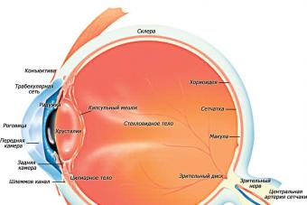
 Let's try to imagine what is inside the eyeball. The optical structure of the eye consists of refractive, accommodative and receptor apparatus.. The following is a brief description of the entire path traveled by a light beam entering the eye. The device of the eyeball in section and the passage of light rays through it will present you with the following figure with symbols.
Cornea
The first eye "lens" on which the beam reflected from the object falls and is refracted is the cornea. This is what the entire optical mechanism of the eye is covered on the front side.
It is she who provides an extensive field of view and clarity of the image on the retina.
Damage to the cornea leads to tunnel vision - a person sees the world around him as if through a pipe. Through the cornea of the eye "breathes" - it passes oxygen from the outside.
Cornea properties:
- Absence of blood vessels;
- Full transparency;
- High sensitivity to external influences.
 The spherical surface of the cornea preliminarily collects all the rays at one point, so that then project it onto the retina. In the likeness of this natural optical mechanism, various microscopes and cameras have been created.
Iris with pupil
Some of the rays that pass through the cornea are filtered out by the iris. The latter is delimited from the cornea by a small cavity filled with a transparent chamber fluid - the anterior chamber.
The iris is a movable opaque diaphragm that regulates the flow of light passing through. The round colored iris is located just behind the cornea.
Its color varies from light blue to dark brown and depends on the race of the person and on heredity.
Sometimes there are people who have left and right eye have a different color. The red color of the iris occurs in albinos.
R  the arcuate membrane is supplied with blood vessels and is equipped with special muscles - annular and radial. The first (sphincters), contracting, automatically narrow the lumen of the pupil, and the second (dilators), contracting, expand it if necessary.
The pupil is located in the center of the iris and is a round hole with a diameter of 2-8 mm. Its narrowing and expansion occurs involuntarily and is not controlled by a person in any way. By narrowing in the sun, the pupil protects the retina from burns. Except from bright light, the pupil constricts from irritation of the trigeminal nerve and from certain medications. Pupil dilation can occur from strong negative emotions (horror, pain, anger).
lens
Further, the light flux enters a biconvex elastic lens - the lens. It is an accommodation mechanism located behind the pupil and delimits the anterior part of the eyeball, including the cornea, iris and anterior chamber of the eye. Behind it tightly adjoins the vitreous body.
 In the transparent protein substance of the lens, there are no blood vessels and innervation. The substance of the organ is enclosed in a dense capsule. The lens capsule is radially attached to the ciliary body of the eye. with the help of the so-called ciliary girdle. Tensioning or loosening this band changes the curvature of the lens, which allows you to clearly see both close and distant objects. This property is called accommodation.
The thickness of the lens varies from 3 to 6 mm, the diameter depends on age, reaching 1 cm in an adult. Newborns and infants are characterized by an almost spherical shape of the lens due to its small diameter, but as the child grows older, the diameter of the lens gradually increases. In older people, the accommodative functions of the eyes deteriorate.
Pathological clouding of the lens is called a cataract.
vitreous body
The vitreous body fills the cavity between the lens and the retina. Its composition is represented by a transparent gelatinous substance that freely transmits light. With age, as well as with high and medium myopia, small opacities appear in the vitreous body, perceived by a person as “flying flies”. The vitreous body lacks blood vessels and nerves.
Retina and optic nerve
After passing through the cornea, pupil and lens, the light rays are focused on the retina. The retina is the inner shell of the eye, characterized by the complexity of its structure and consisting mainly of nerve cells. It is a part of the brain that has grown forward.
The light-sensitive elements of the retina are in the form of cones and rods. The first are the organ of daytime vision, and the second - twilight.
Rods are able to perceive very weak light signals.
Deficiency in the body of vitamin A, which is part of the visual substance of the rods, leads to night blindness - a person does not see well at dusk.
 From the cells of the retina originates the optic nerve, which is connected together nerve fibers emanating from the retina. The place where the optic nerve enters the retina is called the blind spot. since it does not contain photoreceptors. The zone with the largest number of photosensitive cells is located above the blind spot, approximately opposite the pupil, and is called the Yellow Spot.
The human organs of vision are arranged in such a way that on their way to the hemispheres of the brain, part of the fibers of the optic nerves of the left and right eyes intersect. Therefore, in each of the two hemispheres of the brain there are nerve fibers of both the right and left eyes. The point where the optic nerves cross is called the chiasm. The picture below shows the location of the chiasm, the base of the brain.
The construction of the path of the light flux is such that the object viewed by a person is displayed upside down on the retina.
After that, the image is transmitted with the help of the optic nerve to the brain, "turning" it into a normal position. The retina and optic nerve are the receptor apparatus of the eye.
The eye is one of the most perfect and complex creations of nature. The slightest disturbance in at least one of its systems leads to visual disturbances.
Videos that will interest you:
Vision is a biological process that determines the perception of the shape, size, color of objects around us, orientation among them. It is possible due to the function of the visual analyzer, which includes the perceiving apparatus - the eye.
vision function not only in the perception of light rays. We use it to assess the distance, volume of objects, visual perception of the surrounding reality.
Human eye - photo
Currently, of all the sense organs in humans, the greatest load falls on the organs of vision. This is due to reading, writing, watching television and other types of information and work.
The structure of the human eye
The organ of vision consists of the eyeball and an auxiliary apparatus located in the eye socket - a deepening of the bones of the facial skull.
The structure of the eyeball
The eyeball has the appearance of a spherical body and consists of three shells:
- External - fibrous;
- medium - vascular;
- internal - mesh.

Outer fibrous sheath in the posterior part it forms a protein, or sclera, and in front it passes into a cornea permeable to light.
Middle choroid It is called so because of the fact that it is rich in blood vessels. Located under the sclera. The anterior part of this shell forms iris, or the iris. So it is called because of the color (the color of the rainbow). In the iris is pupil- a round hole that is able to change its value depending on the intensity of illumination through an innate reflex. To do this, there are muscles in the iris that narrow and expand the pupil.
The iris acts as a diaphragm that regulates the amount of light entering the light-sensitive apparatus and protects it from damage by accustoming the organ of vision to the intensity of light and darkness. The choroid forms a liquid - the moisture of the chambers of the eye.
Inner retina, or retina- adjacent to the back of the middle (vascular) membrane. Consists of two sheets: outer and inner. The outer sheet contains pigment, the inner sheet contains photosensitive elements.

The retina lines the bottom of the eye. If you look at it from the side of the pupil, then a whitish round spot is visible at the bottom. This is the exit site of the optic nerve. There are no photosensitive elements and therefore no light rays are perceived, it is called blind spot. To the side of it is yellow spot (macula). This is the place of greatest visual acuity.
In the inner layer of the retina are light-sensitive elements - visual cells. Their ends look like rods and cones. sticks contain a visual pigment - rhodopsin, cones- iodopsin. Rods perceive light in twilight conditions, and cones perceive colors in sufficiently bright light.
Sequence of light passing through the eye
Consider the path of light rays through that part of the eye that makes up its optical apparatus. First, the light passes through the cornea, the aqueous humor of the anterior chamber of the eye (between the cornea and the pupil), the pupil, the lens (in the form of a biconvex lens), the vitreous body (a thick, transparent medium) and finally enters the retina.

In cases where light rays, having passed through the optical media of the eye, are not focused on the retina, visual anomalies develop:
- If ahead of her - myopia;
- if behind - farsightedness.
To equalize myopia, biconcave lenses are used, and hyperopia - biconvex lenses.
As already noted, rods and cones are located in the retina. When light hits them, it causes irritation: complex photochemical, electrical, ionic and enzymatic processes occur that cause nervous excitation - a signal. It enters through the optic nerve to the subcortical (quadrigemina, optic tubercle, etc.) centers of vision. Then it goes to the cortex of the occipital lobes of the brain, where it is perceived as a visual sensation.
The entire complex of the nervous system, including light receptors, optic nerves, vision centers in the brain, constitutes the visual analyzer.
The structure of the auxiliary apparatus of the eye

In addition to the eyeball, an auxiliary apparatus also belongs to the eye. It consists of eyelids, six muscles that move the eyeball. The back surface of the eyelids is covered by a shell - the conjunctiva, which partially passes to the eyeball. In addition, the lacrimal apparatus belongs to the auxiliary organs of the eye. It consists of the lacrimal gland, lacrimal ducts, sac and nasolacrimal duct.
The lacrimal gland secretes a secret - tears containing lysozyme, which has a detrimental effect on microorganisms. It is located in the fossa of the frontal bone. Its 5-12 tubules open into the gap between the conjunctiva and the eyeball in the outer corner of the eye. Moisturizing the surface of the eyeball, tears flow to the inner corner of the eye (nose). Here they gather in the openings of the lacrimal ducts, through which they enter the lacrimal sac, also located at the inner corner of the eye.
From the sac along the nasolacrimal duct, tears are directed into the nasal cavity, under the lower concha (therefore, sometimes you can notice how tears flow from the nose while crying).
Vision hygiene
Knowing the ways of outflow of tears from the places of formation - the lacrimal glands - allows you to correctly perform such a hygiene skill as “wiping” the eyes. At the same time, the movement of hands with a clean napkin (preferably sterile) should be directed from the outer corner of the eye to the inner one, “wipe your eyes towards the nose”, towards the natural flow of tears, and not against it, thus contributing to the removal of a foreign body (dust) on the surface of the eyeball.
The organ of vision must be protected from foreign bodies and damage. When working, where particles, fragments of materials, chips are formed, protective glasses should be used.
If vision deteriorates, do not hesitate and contact an ophthalmologist, follow his recommendations in order to avoid further development of the disease. The intensity of lighting in the workplace should depend on the type of work being done: the more subtle movements are performed, the more intense the lighting should be. It should not be bright or weak, but exactly the one that requires the least eye strain and contributes to efficient work.
How to maintain visual acuity
Lighting standards have been developed depending on the purpose of the premises, on the type of activity. The amount of light is determined using a special device - a luxmeter. Control of the correctness of lighting is carried out by the medical and sanitary service and the administration of institutions and enterprises.
It should be remembered that bright light especially contributes to the deterioration of visual acuity. Therefore, you should avoid looking without light-protective glasses towards sources of bright light, both artificial and natural.
To prevent visual impairment due to high eye strain, certain rules must be followed:
- When reading and writing, uniform sufficient lighting is necessary, from which fatigue does not develop;
- the distance from the eyes to the subject of reading, writing or small items with which you are busy should be about 30-35cm;
- the objects with which you work should be placed conveniently for the eyes;
- Watch TV shows no closer than 1.5 meters from the screen. In this case, it is necessary to highlight the room due to a hidden light source.
Of no small importance for maintaining normal vision is a fortified diet in general, and especially vitamin A, which is abundant in animal products, in carrots, pumpkins.
A measured lifestyle, which includes the correct alternation of the regime of work and rest, nutrition, excluding bad habits, including smoking and drinking alcoholic beverages, to a large extent contributes to the preservation of vision and health in general.
Hygienic requirements for the preservation of the organ of vision are so extensive and varied that the above cannot be limited. They may vary depending on the work activity, they should be clarified with a doctor and performed.
We advise you to read
 What is the highest heel in the world?
 Top 10 most educated countries in the world with high literacy rates Countries with the best education in the world
 Fortune telling on playing cards for a loved one Fortune telling for a beloved man on cards 36
 The most educated countries in the world Japan is the most educated country in the world