In the image and likeness ...
Our eye is one of the most important sense organs. Thanks to him, we have access to 90 percent of the information about the entire world around us. In terms of capabilities, it can be compared with a camera. Although, of course, this camera is made in the image and likeness of our eye.
Features of the external structure of the human eye
The eye lies in a kind of "mink", which is called the eye socket.
It is round like an apple, the organ of vision and got its name - "eyeball". It peeks through the gap located between the lower and upper eyelids. The most important feature of the external structure of the eye is a kind of black spot of non-fixed size. This is the pupil. Thanks to him, we, in fact, see the world around us. It can expand and contract. In a dark room, our pupils always dilate to let in as much light as possible into the eyeball, and as soon as we turn on the bright light of the lamp, they immediately decrease, turning from a speck into a dot. Such a funny transformation of the pupil occurs due to the muscle located in the iris of the eye - a colored ring encircling it. Do you know why our pupils are black? Because inside the eye itself - emptiness! Let's move on to its internal structure.
human eye anatomy
On its back and round wall, as on the film of old cameras, there is a whole layer of light-sensitive cells - the retina. She, like a net, catches the rays of light. Imagine, it contains approximately 140 million light-sensitive cells! If a beam of light hits them, then chemical reactions begin, instantly transforming into nerve impulses.

A special optic nerve delivers these impulses to the visual part of the brain, and that, processing the signal, “shows” us a picture. The structure of the human eye is such that the image that our brain shows is exactly the opposite of the image located on the retina. It is the brain that shows us everything in a three-dimensional image, not a flat one. And the brain also “remembers” the distance between the objects we look at. For example, a huge cat and a tiny bus that rushes along the road are objects located at a great distance from each other. Naturally, their true sizes will be directly opposite! One of the structural features of the eye is the lens. He is responsible for a clear image of the type of camera lens.

In fact, this is the lens, only biconvex. In addition, this "lens" is not hard, but elastic.
The lens as a structural feature of the eye
The lens of the eye collects light rays and sends them to the retina. If the object we are looking at is located far from us, then the lens for focusing its (object) rays should become flat, and if we look at close objects - convex. In this case, the muscle that is located around the lens is connected. Having contracted, it makes it flat, relaxing - convex. Imagine with what precision this muscle must work, provided that for all objects located at different distances from each other, different curvature of the lens is required.
The human organ of vision almost does not differ in its structure from the eyes of other mammals, which means that in the process of evolution the structure of the human eye has not undergone significant changes. And today the eye can rightly be called one of the most complex and high-precision devices, created by nature for the human body. You will learn more about how the human visual apparatus works, what the eye consists of and how it works, in this review.
General information about the structure and operation of the organ of vision
The anatomy of the eye includes its external (visually visible from the outside) and internal (located inside the skull) structure. The outer part of the eye that can be seen includes the following bodies:
- eye socket;
- Eyelid;
- Lacrimal glands;
- Conjunctiva;
- Cornea;
- Sclera;
- Iris;
- Pupil.
Outwardly, the eye looks like a slit on the face, but in fact the eyeball has the shape of a ball, slightly elongated from the forehead to the back of the head (along the sagittal direction) and having a mass of about 7 g. farsightedness.
Eyelids, lacrimal glands and eyelashes
These organs do not belong to the structure of the eye, but normal visual function is impossible without them, so they should also be considered. The job of the eyelids is to moisten the eyes, remove debris from them and protect them from injury.
 Regular moistening of the surface of the eyeball occurs when blinking. On average, a person blinks 15 times per minute, while reading or working with a computer - less often. The lacrimal glands, located in the upper outer corners of the eyelids, work continuously, releasing the fluid of the same name into the conjunctival sac. Excess tears are removed from the eyes through the nasal cavity, entering it through special tubules. In a pathology called dacryocystitis, the corner of the eye cannot communicate with the nose due to blockage of the lacrimal canal.
The inner side of the eyelid and the front visible surface of the eyeball is covered with the thinnest transparent membrane - the conjunctiva. It also contains additional small lacrimal glands.
It is its inflammation or damage that causes us to feel sand in the eye.
The eyelid keeps a semicircular shape due to the internal dense cartilaginous layer and circular muscles - palpebral fissures. The edges of the eyelids are decorated with 1-2 rows of eyelashes - they protect the eyes from dust and sweat. Here, the excretory ducts of the small sebaceous glands open, the inflammation of which is called barley.
oculomotor muscles

 These muscles work more actively than all other muscles of the human body and serve to give direction to the gaze. From the inconsistency in the work of the muscles of the right and left eyes, strabismus occurs. Special muscles set the eyelids in motion - raise and lower them. oculomotor muscles are attached with their tendons to the surface of the sclera.
Optical system of the eye
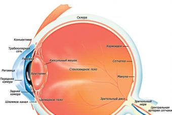
 Let's try to imagine what is inside the eyeball. The optical structure of the eye consists of refractive, accommodative and receptor apparatus.. The following is a brief description of the entire path traveled by a light beam entering the eye. The device of the eyeball in section and the passage of light rays through it will present you with the following figure with symbols.
Cornea
The first eye "lens" on which the beam reflected from the object falls and is refracted is the cornea. This is what the entire optical mechanism of the eye is covered on the front side.
It is she who provides an extensive field of view and clarity of the image on the retina.
Damage to the cornea leads to tunnel vision - a person sees the world around him as if through a pipe. Through the cornea of the eye "breathes" - it passes oxygen from the outside.
Cornea properties:
- Absence of blood vessels;
- Full transparency;
- High sensitivity to external influences.
 The spherical surface of the cornea preliminarily collects all the rays at one point, so that then project it onto the retina. In the likeness of this natural optical mechanism, various microscopes and cameras have been created.
Iris with pupil
Some of the rays that pass through the cornea are filtered out by the iris. The latter is delimited from the cornea by a small cavity filled with a transparent chamber fluid - the anterior chamber.
The iris is a movable opaque diaphragm that regulates the flow of light passing through. The round colored iris is located just behind the cornea.
Its color varies from light blue to dark brown and depends on the race of the person and on heredity.
Sometimes there are people who have left and right eye have a different color. The red color of the iris occurs in albinos.
R  the arcuate membrane is supplied with blood vessels and is equipped with special muscles - annular and radial. The first (sphincters), contracting, automatically narrow the lumen of the pupil, and the second (dilators), contracting, expand it if necessary.
The pupil is located in the center of the iris and is a round hole with a diameter of 2-8 mm. Its narrowing and expansion occurs involuntarily and is not controlled by a person in any way. By narrowing in the sun, the pupil protects the retina from burns. Except from bright light, the pupil constricts from irritation of the trigeminal nerve and from certain medications. Pupil dilation can occur from strong negative emotions (horror, pain, anger).
lens
Further, the light flux enters a biconvex elastic lens - the lens. It is an accommodation mechanism located behind the pupil and delimits the anterior part of the eyeball, including the cornea, iris and anterior chamber of the eye. Behind it tightly adjoins the vitreous body.
 In the transparent protein substance of the lens, there are no blood vessels and innervation. The substance of the organ is enclosed in a dense capsule. The lens capsule is radially attached to the ciliary body of the eye. with the help of the so-called ciliary girdle. Tensioning or loosening this band changes the curvature of the lens, which allows you to clearly see both close and distant objects. This property is called accommodation.
The thickness of the lens varies from 3 to 6 mm, the diameter depends on age, reaching 1 cm in an adult. Newborns and infants are characterized by an almost spherical shape of the lens due to its small diameter, but as the child grows older, the diameter of the lens gradually increases. In older people, the accommodative functions of the eyes deteriorate.
Pathological clouding of the lens is called a cataract.
vitreous body
The vitreous body fills the cavity between the lens and the retina. Its composition is represented by a transparent gelatinous substance that freely transmits light. With age, as well as with high and medium myopia, small opacities appear in the vitreous body, perceived by a person as “flying flies”. The vitreous body lacks blood vessels and nerves.
Retina and optic nerve
After passing through the cornea, pupil and lens, the light rays are focused on the retina. The retina is the inner shell of the eye, characterized by the complexity of its structure and consisting mainly of nerve cells. It is a part of the brain that has grown forward.
The light-sensitive elements of the retina are in the form of cones and rods. The first are the organ of daytime vision, and the second - twilight.
Rods are able to perceive very weak light signals.
Deficiency in the body of vitamin A, which is part of the visual substance of the rods, leads to night blindness - a person does not see well at dusk.
 From the cells of the retina originates the optic nerve, which is connected together nerve fibers emanating from the retina. The place where the optic nerve enters the retina is called the blind spot. since it does not contain photoreceptors. The zone with the largest number of photosensitive cells is located above the blind spot, approximately opposite the pupil, and is called the Yellow Spot.
The human organs of vision are arranged in such a way that on their way to the hemispheres of the brain, part of the fibers of the optic nerves of the left and right eyes intersect. Therefore, in each of the two hemispheres of the brain there are nerve fibers of both the right and left eyes. The point where the optic nerves cross is called the chiasm. The picture below shows the location of the chiasm, the base of the brain.
The construction of the path of the light flux is such that the object viewed by a person is displayed upside down on the retina.
After that, the image is transmitted with the help of the optic nerve to the brain, "turning" it into a normal position. The retina and optic nerve are the receptor apparatus of the eye.
The eye is one of the most perfect and complex creations of nature. The slightest disturbance in at least one of its systems leads to visual disturbances.
Videos that will interest you:
A person sees not with his eyes, but through his eyes, from where information is transmitted through the optic nerve, chiasm, visual tracts to certain areas of the occipital lobes of the cerebral cortex, where the picture of the outside world that we see is formed. All these organs make up our visual analyzer or visual system.
The presence of two eyes allows us to make our vision stereoscopic (that is, to form a three-dimensional image). The right side of the retina of each eye transmits through the optic nerve the "right side" of the image to the right side of the brain, the left side of the retina does the same. Then the two parts of the image - the right and left - the brain connects together.
Since each eye perceives “its own” picture, if the joint movement of the right and left eyes is disturbed, binocular vision can be upset. Simply put, you will begin to see double, or you will see two completely different pictures at the same time.
Basic functions of the eye
- an optical system that projects an image;
- a system that perceives and “encodes” the received information for the brain;
- "serving" life support system.
The eye can be called a complex optical device. Its main task is to “transmit” the correct image to the optic nerve.
Cornea- a transparent membrane that covers the front of the eye. There are no blood vessels in it, it has a large refractive power. Included in the optical system of the eye. The cornea borders on the opaque outer shell of the eye - the sclera. See the structure of the cornea.
Anterior chamber of the eye is the space between the cornea and the iris. It is filled with intraocular fluid.
iris- in shape it is similar to a circle with a hole inside (pupil). The iris consists of muscles, with the contraction and relaxation of which the size of the pupil changes. It enters the choroid of the eye. The iris is responsible for the color of the eyes (if it is blue, it means that there are few pigment cells in it, if it is brown, there are many). It performs the same function as the aperture in a camera, adjusting the light output.
Pupil- a hole in the iris. Its dimensions usually depend on the level of illumination. The more light, the smaller the pupil.
lens- "natural lens" of the eye. It is transparent, elastic - it can change its shape, “focusing” almost instantly, due to which a person sees well both near and far. Enclosed in a capsule ciliary girdle. The lens, like the cornea, is part of the optical system of the eye.
vitreous body- a gel-like transparent substance located in the back of the eye. The vitreous body maintains the shape of the eyeball and is involved in intraocular metabolism. Included in the optical system of the eye.
Retina- consists of photoreceptors (they are sensitive to light) and nerve cells. Receptor cells located in the retina are divided into two types: cones and rods. In these cells, which produce the enzyme rhodopsin, the energy of light (photons) is converted into electrical energy of the nervous tissue, i.e., a photochemical reaction.
The rods are highly sensitive to light and allow you to see in low light, they are also responsible for peripheral vision. Cones, on the contrary, require more light for their work, but it is they that allow you to see fine details (are responsible for central vision), make it possible to distinguish colors. The greatest concentration of cones is in the fovea (macula), which is responsible for the highest visual acuity. The retina is adjacent to the choroid, but loosely in many areas. It is here that it tends to flake off in various diseases of the retina.
Sclera- an opaque outer shell of the eyeball, passing in front of the eyeball into a transparent cornea. 6 oculomotor muscles are attached to the sclera. It contains a small number of nerve endings and blood vessels.
choroid- lines the posterior sclera, adjacent to the retina, with which it is closely connected. The choroid is responsible for the blood supply to the intraocular structures. In diseases of the retina, it is very often involved in the pathological process. There are no nerve endings in the choroid, therefore, when it is ill, pain does not occur, usually signaling some kind of malfunction.
optic nerve- With the help of the optic nerve, signals from the nerve endings are transmitted to the brain.
The visual organs of fish are basically the same as those of other vertebrates. The mechanism of perception of visual sensations is similar to other vertebrates: light passes into the eye through the transparent cornea, then the pupil - a hole in the iris - passes it to the lens, and the lens transmits and focuses the light on the inner wall of the eye to the retina, where it is directly perceived. . The retina consists of light-sensitive (photoreceptor), nerve, as well as supporting cells.
Light-sensitive cells are located on the side of the pigment membrane. In their processes, shaped like rods and cones, there is a photosensitive pigment. The number of these photoreceptor cells is very large - there are 50 thousand of them per 1 mm 2 of the retina in carp (in squid - 162 thousand, spider - 16 thousand, human - 400 thousand, owl - 680 thousand). Through a complex system of contacts between the terminal branches of sensory cells and dendrites of nerve cells, light stimuli enter the optic nerve.
Cones in bright light perceive the details of objects and color. Rods perceive weak light, but they cannot create a detailed image.
The position and interaction of the cells of the pigment membrane, rods and cones change depending on the illumination. In the light, the pigment cells expand and cover the rods located near them; cones are drawn to the nuclei of cells and thus move towards the light. In the dark, sticks are drawn to the nuclei (and are closer to the surface); the cones approach the pigment layer, and the pigment cells reduced in the dark cover them.
The number of receptors of various kinds depends on the way of life of fish. In diurnal fish, cones prevail in the retina, in twilight and nocturnal fish, rods: burbot has 14 times more rods than pike. Deep-sea fish living in the darkness of the depths do not have cones, and the rods become larger and their number increases sharply - up to 25 million / mm 2 of the retina; the probability of capturing even weak light increases. Most fish distinguish colors, which is confirmed by the possibility of developing conditioned reflexes in them for a certain color - blue, green, red, yellow, blue.
Some deviations from the general scheme of the structure of the eye of a fish are associated with the characteristics of life in the water. The eye of the fish is elliptical. Among others, it has a silvery shell (between the vascular and protein), rich in guanine crystals, which gives the eye a greenish-golden sheen.
The cornea is almost flat (rather than convex), the lens is spherical (rather than biconvex) - this expands the field of view. The hole in the iris - the pupil - can change the diameter only within small limits. As a rule, fish do not have eyelids. Only sharks have a nictitating membrane that covers the eye like a curtain, and some herring and mullet have a fatty eyelid - a transparent film covering part of the eye.
The location of the eyes on the sides of the head (in most species) is the reason why fish have mostly monocular vision, and the ability for binocular vision is very limited. The spherical shape of the lens and moving it forward to the cornea provides a wide field of view: light enters the eye from all sides. The vertical angle of view is 150°, horizontally 168–170°. But at the same time, the sphericity of the lens causes myopia in fish. The range of their vision is limited and fluctuates due to the turbidity of the water from a few centimeters to several tens of meters.
Vision over long distances becomes possible due to the fact that the lens can be pulled back by a special muscle - a sickle-shaped process extending from the choroid of the bottom of the eyecup.
With the help of vision, fish are also guided by objects on the ground. Improved vision in the dark is achieved by the presence of a reflective layer (tapetum) - guanine crystals, underlain by pigment. This layer does not transmit light to the tissues lying behind the retina, but reflects it and returns it back to the retina. This increases the ability of the receptors to use the light that has entered the eye.
Due to habitat conditions, the eyes of fish can change greatly. In cave or abyssal (deep water) forms, the eyes can be reduced and even disappear. Some deep-sea fish, on the contrary, have huge eyes that allow them to capture very faint traces of light, or telescopic eyes, the collecting lenses of which the fish can put in parallel and acquire binocular vision. The eyes of some eels and larvae of a number of tropical fish are carried forward on long outgrowths (stalked eyes).
An unusual modification of the eyes of a four-eyed bird from Central and South America. Her eyes are placed on the top of her head, each of them is divided by a partition into two independent parts: the upper fish sees in the air, the lower one in the water. In the air, the eyes of fish crawling ashore or trees can function.
The role of vision as a source of information from the outside world for most fish is very large: when orienting during movement, when searching for and capturing food, while maintaining a flock, during the spawning period (the perception of defensive and aggressive postures and movements by rival males, and between individuals of different sexes - wedding attire and spawning "ceremonial"), in the relationship of the victim-predator, etc.
The ability of fish to perceive light has long been used in fishing (fishing by the light of a torch, fire, etc.).
It is known that fish of different species react differently to light of different intensities and different wavelengths, i.e., different colors. Thus, bright artificial light attracts some fish (Caspian sprat, saury, horse mackerel, mackerel, etc.) and scares away others (mullet, lamprey, eel, etc.). Different species are also selectively related to different colors and different light sources - surface and underwater. All this is the basis for the organization of industrial fishing for electric light (this is how sprat, saury and other fish are caught).

The eye apparatus is stereoscopic and in the body is responsible for the correct perception of information, the accuracy of its processing and further transmission to the brain.
The right side of the retina sends information from the right lobe of the image to the brain through transmission through the optic nerve, the left side transmits the left lobe, as a result, the brain connects both, and a common visual picture is obtained.
The lens is fixed with thin threads, one end of which is tightly woven into the lens, its capsule, and the other end is connected to the ciliary body.
When the tension of the threads changes, the process of accommodation occurs . The lens is devoid of lymphatic vessels and blood vessels, as well as nerves.
It provides the eye with light transmission and refraction, endows it with the function of accommodation, and is the eye's divider into the posterior and anterior regions.
vitreous body
The vitreous body of the eye is the largest formation. This is a colorless substance of a gel-like substance, which is formed in the form of a spherical shape, in the sagittal direction it is flattened.
The vitreous body consists of a gel-like substance of organic origin, a membrane and a vitreous canal.
In front of it is the lens, zonular ligament and ciliary processes, its back part comes close to the retina. The connection of the vitreous body and the retina occurs at the optic nerve and in the part of the dentate line, where the flat part of the ciliary body is located. This area is the base of the vitreous body, and the width of this belt is 2-2.5 mm.
The chemical composition of the vitreous body: 98.8 hydrophilic gel, 1.12% dry residue. When a hemorrhage occurs, the thromboplastic activity of the vitreous body increases dramatically.
This feature is aimed at stopping bleeding. In the normal state of the vitreous body, fibrinolytic activity is absent.
Nutrition and maintenance of the vitreous body environment is provided by the diffusion of nutrients that through the vitreous membrane enter the body from the intraocular fluid and osmosis.
There are no vessels and nerves in the vitreous body, and its biomicroscopic structure presents various forms of gray ribbons with white speckles. Between the ribbons there are areas without color, completely transparent.
Vacuoles and opacities in the vitreous body appear with age. In the case when there is a partial loss of the vitreous body, the place is filled with intraocular fluid.
Chambers with aqueous humor
The eye has two chambers that are filled with aqueous humor. Moisture is formed from the blood by processes of the ciliary body. Its release occurs first in the anterior chamber, then it enters the anterior chamber.
Aqueous moisture enters the anterior chamber through the pupil. The human eye produces 3 to 9 ml of moisture per day. Aqueous moisture contains substances that nourish the lens, the corneal endothelium, the anterior vitreous, and the trabecular meshwork.
It contains immunoglobulins that help remove dangerous factors from the eye, its inner part. If the outflow of aqueous humor is impaired, then this can develop an eye disease such as glaucoma, as well as an increase in pressure inside the eye.
In cases of violation of the integrity of the eyeball, the loss of aqueous humor leads to hypotension of the eye.
Iris

The iris is the avant-garde part of the vascular tract. It is located just behind the cornea, between the chambers and in front of the lens. The iris is round in shape and is located around the pupil.
It consists of a boundary layer, a stromal layer, and a pigment-muscle layer. It has an uneven surface with a pattern. The iris contains pigment cells, which are responsible for the color of the eyes.
The main tasks of the iris: regulation of the light flux that passes to the retina through the pupil and protection of light-sensitive cells. Visual acuity depends on the correct functioning of the iris.
The iris has two muscle groups. One group of muscles is deployed around the pupil and regulates its reduction, the other group is deployed radially along the thickness of the iris, regulating the expansion of the pupil. The iris has many blood vessels.
Retina
It is an optimally thin shell of the nervous tissue and represents the peripheral part of the visual analyzer. There are photoreceptor cells in the retina, which are responsible for perception, as well as for converting electromagnetic radiation into nerve impulses. It is adjacent from the inside to the vitreous body, and to the vascular layer of the eyeball - from the outside.

The retina has two parts. One part is visual, the other is the blind part, which does not contain photosensitive cells. The internal structure of the retina is divided into 10 layers.
The main task of the retina is to receive the light flux, process it, converting it into a signal that forms complete and encoded information about the visual image.
optic nerve
The optic nerve is a network of nerve fibers. Among these thin fibers is the central canal of the retina. The starting point of the optic nerve is located in the ganglion cells, then its formation occurs by passing through the sclera membrane and fouling of the nerve fibers with meningeal structures.
The optic nerve has three layers - hard, arachnoid, soft. There is liquid between the layers. The diameter of the optic disc is about 2 mm.
Topographic structure of the optic nerve:
- intraocular;
- intraorbital;
- intracranial;
- intratubular;
How the human eye works
The light flux passes through the pupil and through the lens is brought into focus on the retina. The retina is rich in light-sensitive rods and cones, of which there are more than 100 million in the human eye.
Video: "The process of vision"
The rods provide sensitivity to light, and the cones give the eyes the ability to see colors and small details. After the refraction of the light flux, the retina transforms the image into nerve impulses. Further, these impulses pass to the brain, which processes the information received.
Diseases
Diseases associated with a violation of the structure of the eye can be caused both by an incorrect arrangement of its parts in relation to each other, and by internal defects in these parts.
The first group includes diseases that lead to a decrease in visual acuity:
- Myopia. It is characterized by an increased length of the eyeball compared to the norm. This causes the light passing through the lens to be focused not on the retina, but in front of it. The ability to see objects at a distance from the eyes is impaired. Myopia corresponds to a negative number of diopters when measuring visual acuity.
- Farsightedness. It is a consequence of a decrease in the length of the eyeball or loss of elasticity of the lens. In both cases, the accommodative possibilities are reduced, the correct focusing of the image is disturbed, and the light rays converge behind the retina. The ability to see nearby objects is impaired. Farsightedness corresponds to a positive number of diopters.
- Astigmatism. This disease is characterized by a violation of the sphericity of the eye membrane due to defects in the lens or cornea. This leads to an uneven convergence of the rays of light entering the eye, the clarity of the image received by the brain is disturbed. Astigmatism is often accompanied by nearsightedness or farsightedness.
Pathologies associated with functional disorders of certain parts of the organ of vision:
- Cataract. With this disease, the lens of the eye becomes cloudy, its transparency and ability to conduct light are disturbed. Depending on the degree of clouding, visual impairment can be different up to complete blindness. Most people develop cataracts in old age but do not progress to severe stages.
- Glaucoma is a pathological change in intraocular pressure. It can be provoked by many factors, for example, a decrease in the anterior chamber of the eye or the development of cataracts.
- Myodesopsia or "flying flies" before the eyes. It is characterized by the appearance of black dots in the field of view, which can be presented in different quantities and sizes. Points arise due to violations in the structure of the vitreous body. But in this disease, the causes are not always physiological - “flies” can appear due to overwork or after suffering infectious diseases.
- Strabismus. It is provoked by a change in the correct position of the eyeball in relation to the eye muscle or a violation of the work of the eye muscles.
- Retinal detachment. The retina and posterior vascular wall are separated from each other. This is due to a violation of the tightness of the retina, which occurs when its tissues break. Detachment is manifested by clouding of the outline of objects before the eyes, the appearance of flashes in the form of sparks. If some corners fall out of the field of view, this means that the detachment has taken severe forms. If left untreated, complete blindness occurs.
- Anophthalmos - underdevelopment of the eyeball. A rare congenital pathology, the cause of which is a violation of the formation of the frontal lobes of the brain. Anophthalmos can also be acquired, then it develops after surgical operations (for example, to remove tumors) or severe eye injuries.
Prevention
- You should take care of the health of the circulatory system, especially that part of it that is responsible for the flow of blood to the head. Many visual defects are due to atrophy and damage to the ophthalmic and brain nerves.
- Eye strain must not be allowed. When working with constant examination of small objects, you need to take regular breaks with eye exercises. The workplace should be equipped so that the brightness of the lighting and the distance between objects are optimal.
- The intake of a sufficient amount of minerals and vitamins in the body is another condition for maintaining healthy vision. Vitamins C, E, A and minerals such as zinc are especially important for the eyes.
- Proper eye hygiene helps prevent the development of inflammatory processes, the complications of which can significantly impair vision.
Bibliography
- Ophthalmology. National leadership. Short edition Ed. S.E. Avetisova, E.A. Egorova, L.K. Moshetova, V.V. Neroeva, Kh.P. Tahchidi 2019
- Atlas of ophthalmology G.K. Kriglstein, K.P. Ionescu-Cypers, M. Severin, M.A. Wobig 2009
We advise you to read
 What is the highest heel in the world?
 Top 10 most educated countries in the world with high literacy rates Countries with the best education in the world
 Fortune telling on playing cards for a loved one Fortune telling for a beloved man on cards 36
 The most educated countries in the world Japan is the most educated country in the world