One of the most important functions of the human body is movement in space. It is performed by the musculoskeletal system, which consists of two parts: active and passive. Passive bones are connected using various types of joints, active - muscles.
Skeleton(from the Greek. skeletos - dried, dried) is a complex of bones that perform many functions: supporting, protective, locomotor, shaping, overcoming the force of gravity. The total mass of the skeleton is from 1/7 to 1/5 of the mass of the human body. The human skeleton includes more than 200 bones, 33-34 bones of the skeleton are not paired. These are the vertebrae, sacrum, tailbone, some bones of the skull and sternum, the rest of the bones are paired. The skeleton is conventionally divided into two parts: axial and accessory. The axial skeleton includes the vertebral column (26 bones), the skull (29 bones), the chest (25 bones); to the accessory - the bones of the upper (64) and lower (62) limbs.
The bones of the skeleton are the levers that are set in motion by the muscles. As a result, parts of the body change their position in relation to each other and move the body in space. Ligaments, muscles, tendons, fascia are attached to the bones, which are elements of the soft skeleton or soft skeleton, which also takes part in holding the organs near the bones that form a hard (hard) skeleton. The skeleton forms a receptacle for organs, protecting them from external influences: the brain is located in the cranial cavity, the spinal cord is located in the spinal canal, the heart, large vessels, lungs, esophagus, etc., in the pelvic cavity, are located in the pelvic cavity.
Bones represent an unusually complex and very durable complex of spatial systems, which prompted architects to create "perforated structures".
Bones withstand heavy loads... Thus, the tibia can withstand a weight 2 thousand times greater than its weight (1650 kg), the humerus - 850 kg, the tibia - up to 1500 kg.
Bones are involved in mineral metabolism, they are a depot of calcium, phosphorus, etc. Living bone contains vitamins A, Z), C, etc. The vital activity of the bone depends on the functions of the pituitary gland, thyroid and parathyroid glands, adrenal glands and gonads (gonads).
The skeleton is formed by varieties of connective tissue - bone and cartilage, which consist of cells and dense intercellular substance. Bones and cartilage are closely related to each other by a common structure, origin and function. Most bones (bones of the limbs, base of the skull, vertebrae) develop from cartilage, their growth is ensured by proliferation (an increase in the number of cells). A small number of bones develop without the involvement of cartilage (bones of the roof of the skull, lower jaw, collarbone). Some cartilage is not associated with bone and does not change throughout a person's life (cartilage of the auricles, airways). Some cartilage is functionally connected to bone (articular cartilage, menisci).
In the embryo of humans and other vertebrates, the cartilaginous skeleton makes up about 50% of the total body weight. However, gradually cartilage is replaced by bone; in an adult, the mass of cartilage reaches about 2% of the body weight. These are articular cartilage, intervertebral discs, cartilage of the nose and ear, larynx, trachea, bronchi and ribs. Cartilage has the following functions:
- cover articulated surfaces which are therefore highly resistant to wear;
- articular cartilage and intervertebral discs, which are objects of application of compression and stretching forces, carry out their transfer and amortization;
- the cartilages of the airways and outer ear form the walls of the cavities. Muscles, ligaments, tendons are attached to other cartilages.
Cartilage tissue contains about 70-80% water, 10-15 - organic substances, 4-7% salts. Collagen accounts for about 50-70% of the dry matter of cartilage. Depending on the composition, cartilage is hyaline, elastic and collagenous. Like other types of connective tissue, cartilage tissue consists of a few cells (chondrocytes) and the dense intercellular substance they produce. Cartilage does not have blood vessels; it is nourished by diffusion from the surrounding tissues.
Hyaline cartilage smooth, shiny, bluish-white. Basically, the skeleton of the embryo is formed from it, in an adult - the costal cartilage, most of the cartilage of the larynx, the cartilage of the nose, trachea, bronchi and articular (with age, hyaline cartilage becomes calcified).
Elastic cartilage less transparent, yellowish. The auricle, the vocal processes of the arytenoid cartilage of the larynx and the auditory tube are composed of elastic cartilage tissue.
Fibrous cartilage forms intervertebral discs, menisci of the knee and temporomandibular joints. Fibrous cartilage is found in the areas of attachment of ligaments and tendons to bones and cartilage.
Bones are formed by bone tissue, the mechanical properties of which determine the function of the bones. Thus, the tensile resistance of fresh bone and pure copper is the same and is 9 times greater than the resistance of lead. Bone can withstand compression of 10 kg / mm 2 (similar to cast iron), while brick is only 0.5 kg / mm 2. The breaking strength of the ribs is 110 kg / cm 2. This is due to the peculiarities of the chemical composition, structure and architectonics of bones. The bone water content reaches 50%. The dry residue of bone tissue contains about 33% organic and 6-7% inorganic substances.
Bone consists of cells (osteoblasts and osteocytes) and intercellular substance. Osteoblasts are polygonal, cubic, processional young cells, osteocytes are mature multi-procession spindle-shaped cells. Osteoblasts synthesize the components of the intercellular substance and release them from the cell across the entire surface in different directions, which leads to the formation of lacunae (spaces) in which they lie, turning into osteocytes.
Distinguish two types of bone tissue: reticulofibrous (coarsely fibrous) and lamellar. Reticulofibrous bone tissue is located in the areas of attachment of the tendons to the bones, in the seams of the skull after they are overgrown. It consists of thick, disordered bundles of collagen fibers, between which there is an amorphous substance. Osteocytes lie in the gaps.
Lamellar bone tissue is most abundant in the body. It is formed by bone plates with a thickness of 4 to 15 microns, which consist of osteocytes and fine-fibrous bone base material. The fibers forming the laminae lie parallel to each other and are oriented in a specific direction. In this case, the fibers of the adjacent plates are multidirectional and intersect almost at right angles, which provides greater bone strength.
The bone outside, in addition to the articulated surfaces, is covered by the periosteum, which is a strong connective tissue plate rich in blood and lymphatic vessels and nerves. The periosteum is firmly adhered to the bone with the help of connective tissue perforating fibers that penetrate deep into the bone. In the inner layer of the periosteum there are thin spindle-shaped "resting" osteogenic cells, due to which the development, growth in thickness and regeneration of bones after injury occurs.
The bones of a living person- a dynamic structure in which there is a constant metabolism, anabolic and kabolytic processes, destruction of old and creation of new bone plates. Bones adapt to the changing conditions of the body's vital activity, under the influence of which there is a restructuring of their macro- and microscopic structure. The external shape of the bones changes under the influence of stretching and pressure, and bones develop better, the more intensively the activity of the muscles associated with them.
Vertebral column
The vertebral column is composed of 33 separate vertebrae. There are cervical (7 cervical vertebrae), thoracic (12 thoracic), lumbar (5 lumbar), sacral (5 sacral) and coccygeal (4 or 5 coccygeal vertebrae). The sacral and coccygeal vertebrae grow together and form the sacrum and coccyx.
A typical vertebra has a body, a neural arch that surrounds and protects the spinal cord, and seven processes. An unpaired, backward-facing process is called spinous. It serves to attach ligaments and muscles. The vertebral bodies are interconnected with the help of intervertebral cartilage, which, together with the ligaments and muscles along the spine, hold the body in an upright position.
All vertebrae differ in shape and size, especially the first two cervical vertebrae differ from the others - atlas and epistrophy. The movable connection of these vertebrae facilitates the movement of the head. The rest of the vertebrae, the lower they are, the more massive, since they are heavier. The spinal cord is located within the vertebral column in the vertebral canal formed by the holes in the vertebrae. It is reliably protected from all sides.
The vertebral column has bends forward - lordosis, backward (posteriorly) - kephosis, to the sides - scoliosis. The bends of the spinal column increase its spring properties, i.e. promote springy movements of the spinal column. Under the influence of external influences, the bends can change during the day. Therefore, the height of the spine, and consequently, the height of a person can fluctuate during the day on average from 1 to 2-2.5 cm.
The spinal column of a newborn has no bends, they appear during the growth of the body. At the beginning, the newborn develops cervical lordosis (as the child begins to hold his head), then thoracic kephosis (the child begins to sit), and then lumbar lordosis (he begins to stand) and sacral kephosis. By the age of five or six, the bends are clearly visible. In school-age children, severe scoliosis can often be observed.
Rib cage
The rib cage is supported at the back by the spine. On both sides of it, flat bones depart - ribs, representing bony curved plates. In the rib, a middle part (body) and two ends (front and back) are distinguished. The posterior end of the rib has a thickening - the head, which is articulated with the body of the spine by means of a composite surface. Behind the head of the rib is the middle part - the neck, and behind it is the tubercle.
Each rib is articulated with two vertebrae at the same time. The exception is the 9th (not always), 10. And, the 12th thoracic vertebrae, each of which is connected to one rib. The front ends of the ribs are directed towards the sternum. The cartilage of the upper seven pairs of ribs grows to the sternum (true, or pectoral, ribs). The next three pairs of ribs (8, 9, 10th) each grow with their own cartilage to the cartilage of the overlying pair, forming a costal arch. These are the so-called false ribs. The last two pairs (11th, 12th) do not reach the sternum and are very variable in length (free ribs).
Respiratory muscles and a diaphragm are attached to the ribs. When inhaling, the ribs are removed with the front ends from the spine forward and rise upward.
Shoulder girdle
The shoulder girdle consists of two pairs of bones - the shoulder blades and the collarbones. The bones and joints of the shoulder girdle give the arm support and firmly bind it to the torso.
The pelvic girdle is formed by three pairs of bones: the sciatic, pubic, and iliac. The pelvic bones support the entire weight of the trunk.
The skeleton of the upper limbs is formed by: the humerus, the radius and ulna of the forearm, eight small bones of the wrist, five thin metacarpal bones and phalanges of the fingers. Each finger has three phalanges, except for the thumb, which has only two.
The skeleton of the lower extremities consists of the femur (thigh), the tibia and fibula (in the lower leg), 7 bones of the tarsus (in the ankle and heel area), 5 bones of the metatarsus (in the forefoot) and 14 phalanges of the malts.
Scull
The skull has two sections: cerebral and facial. The cerebral skull protects the brain. The bone plates that make up it are very durable. The cranium is formed by the following bones: frontal, two temporal, occipital, two maxillary, two zygomatic, two nasal, vomer, two lacrimal, hyoid bone, palatine. The only movable bone of the skull is the lower jaw.
Some of the bones of the skull are penetrated by sinuses containing air (jaw, frontal, sinuses of the main and ethmoid bones).
This reduces the overall weight of the skull. It is connected to the spine by two occipital condyles.
Bone joints
The joints between the bones of the skull are immobile and strong due to the tight entry of the teeth of one bone into the notches of the other. These joints are called seams. On the contrary, joints are movable joints. For example, the joint between the femur and the pelvic bones, between the humerus and the scapula, resembles a ball-and-socket joint. They are called ball joints. This form makes completely free movements forward and backward, sufficiently wide movements to the sides, rotation inward and outward.
In each the joint has three main elements: articular surfaces, articular capsule and articular cavity. The articular surfaces are covered with cartilage. The articular capsule (bag) is stretched between the articulating bones; it attaches along the edges of the articular surfaces and passes into the periosteum. In the joint capsule, two layers are distinguished: the outer layer is fibrous and the inner layer is synovial. The articular surface is slit-shaped and is located in the articular capsule. The joint cavity contains a small amount of synovial (inter-articular) fluid, which lubricates the articular cartilage, thereby reducing friction in the joints during movement.
Shaped joints are divided into spherical, elliptical, saddle-shaped, block-shaped, flat, etc. Depending on the articular surfaces, some joints can move around one axis (uniaxial joints), in others around two (biaxial joints), in others around three axes (triaxial joints). Uniaxial ones include block and cylindrical. For example, the knee joint is block-rotational in shape, and the ankle joint is block-shaped. A joint is called simple if it is formed by two bones, for example, the humerus, and complex if it is formed by three or more bones.
The skeleton performs not only the musculoskeletal function, but also takes part in the metabolism: it actively participates in maintaining the mineral composition of the blood at a certain level. A number of substances that make up the bone (phosphorus, calcium, citric acid) can enter into metabolic reactions.
Skeleton- the main depot of calcium and phosphorus. The main compound of the mineral component of bone tissue is calcium phosphate. In addition to the basic elements (calcium, phosphorus and magnesium), the bone tissue contains a number of trace elements. Their number is very small, but, nevertheless, they play a large role as biological catalysts for hormones, vitamins and enzymes. Currently, more than 30 trace elements are known that are contained in bone tissue (copper, strontium, zinc, barium, etc.). The content of trace elements in bone tissue varies with age. The accumulation of some of them gradually occurs, which is the reason for the increase in fragility and fragility of the bone with age. These microelements replace calcium ions in the crystal lattice, which leads to the loss of the mechanical strength of the bone.
If calcium is removed from the body more than you eat, a disease of the skeletal system develops in children and adults, which is expressed in changes and curvature of the skeleton in children and softening of bones in adults. A similar disease can develop with low absorption of calcium in the intestine (rickets). The disease is treated with large doses of group vitamins /). Rickets can occur with an excess of certain trace elements in the soil, water and air. So, for example, an excess of beryllium in the soil leads to its excessive accumulation in bone tissue, to the displacement of calcium and to the emergence of "beryllium rickets", which cannot be cured by vitamin D. Excessive intake of aluminum leads to the formation of insoluble aluminum compounds with phosphates in the stomach, as a result, an insufficient amount of phosphorus enters the skeleton.
Normally, two opposite processes continuously occur in bone tissue - reproduction and dissolution of bone substance. At an early age, there is both intensive bone formation and resorption from the side of the medullary canal, therefore, the thickness of the bone walls during this period does not change. By the age of 12, there is a predominance of the process of bone formation and thickening of the bone walls. After a period of stabilization (over 40 years), the process of resorption begins to prevail. The walls of the bone are reduced, they become fragile and easily injured. The change in the mechanical properties of bone is also facilitated by the strong mineralization of osteocytes, which develops with the accumulation of minerals in the bone tissue. Thus, with age, the content of mineral salts increases and the content of the amount of water and organic matter decreases.
In a newborn, the bone contains red bone marrow, the purpose of which is to produce red blood cells (erythrocytes). After birth, the bone marrow, which is located in the cavities of the bone tubes, loses the function of hematopoiesis and becomes yellow bone marrow - an accumulation of intraosseous adipose tissue. But in all flat bones (sternum, etc.) and at the ends of long bones, red bone marrow remains.
The totality of all human bones is called the skeleton, which is the main part of the musculoskeletal system of the body. In this article, we will tell you what kind of tissue the bones are formed by, indicate their number, analyze the varieties by department, and designate the functions of the musculoskeletal system.
general characteristics
The number of bones in the human skeleton depends on age. For example, an adult has about 206, and a child - 270. This difference is due to the fact that some bones of the human skeleton grow together over time (skull, spine, pelvis). In the body, the main part is paired bones, unpaired only 33.
If we talk about the number by department, then:
- the skull consists of 23 bones;
- spine - about 33;
- thoracic region - 25;
- upper limbs - 64;
- lower limbs - 62.

Rice. 1. List of bones.
Each bone organ consists of:
- bone tissue;
- periosteum;
- connecting layer (endost);
- articular cartilage;
- nerves;
- blood vessels.

Rice. 2. Bone structure.
The chemical composition includes mineral salts - 45% (calcium, sodium, potassium, etc.); 25% water; 30% - organic compounds. In addition, this organ is a receptacle of the bone marrow, which performs a hematopoietic function.
The bones of the human skeleton serve as a support for soft tissues, contain and protect internal organs, and participate in metabolic processes. They are formed from bone tissue, which comes from the mesenchyme, and cartilage tissue.
The word "skeleton" of ancient Greek origin and translated - "dried". This is due to the way it is obtained - drying in hot sand or the sun.
Classification
According to their structure and shape, bones are:
TOP-2 articleswho read along with this
- long (shoulder, femoral) - serve to fasten the muscular system of the limbs, act as levers;
- short;
- flat (skull, sternum, ribs, shoulder blades, pelvis) - are the basis of some muscles, protect internal organs;
- airways (skull, face) - consist of air cells and sinuses.

Rice. 3. Varieties of bone organs.
The skeleton does not include six auditory ossicles (three on both sides). They are connected only to each other and carry out the transmission of sound from the eardrum to the inner ear.
Functions
The musculoskeletal system performs biological and mechanical functions.
Biological includes:
- blood-forming - ensures the formation of new blood cells;
- metabolic processes - salt metabolism (the skeleton contains calcium and phosphorus salts).
The mechanical function is:
- support - support of the body, attachment of muscles, internal organs;
- movement - movable joints ensure the work of the bone as a lever, which is set in motion with the help of muscles;
- protection of internal organs;
- shock absorption - structural features soften and reduce shock when moving the body.
What have we learned?
The human skeleton consists of 206 parts, each of which can have its own shape and perform a specific function. The composition of the bone organ is unchanged and includes water, mineral salts and organic matter. With the help of the musculoskeletal system, the human body can move, maintain a constant salt balance, and form new blood cells.
Test by topic
Assessment of the report
Average rating: 4.6. Total ratings received: 956.
In the human body, everything is interconnected and arranged very wisely. The skin and muscles, internal organs and skeleton, all this clearly interacts with each other, thanks to the efforts of nature. Below is a description of the human skeleton and its function.
In contact with
general information
A skeleton made of bones of different sizes and shapes, on which the human body is fixed, is called a skeleton. It serves as a support and provides reliable security for important internal organs. What a human skeleton looks like can be seen in the photo.
Described organ connecting with muscle tissue, it is the musculoskeletal system of homo sapiens. Thanks to this, all individuals can move freely.
The finally developed bone tissue consists of 20% water and is the strongest in the body. Human bones include inorganic substances, thanks to which they have strength, and organic ones, which give flexibility. That is why bones are strong and resilient.
Human Bone Anatomy
 Examining the organ in more detail, it can be seen that it consists of several layers:
- External. Forms high strength bone tissue;
- Connective. The layer tightly covers the outside of the bones;
- Loose connective tissue. Complex interlacing of blood vessels is located here;
- Cartilage tissue. It settled on the ends of the organ, due to it, the bones have the opportunity to grow, but up to a certain age;
- Nerve endings. They carry signals from the brain and vice versa, like wires.
The bone marrow is placed in the cavity of the bone tube; it is red and yellow.
Functions
Without exaggeration, we can say that the body will die if the skeleton ceases to perform its important functions:
- Support... The solid bone-cartilaginous skeleton of the body is formed by the bones, to which fascia, muscles and internal organs are attached.
- Protective... Containers have been created for the maintenance and protection of the spinal cord (spine), the brain (cranium) and for the rest, no less important, organs of human life (rib cage).
- Motor... Here, bones are exploited by muscles as levers to move the body with the help of tendons. They predetermine the coherence of joint movements.
- Cumulative... In the central cavities of the long bones, fat is accumulating - this is the yellow bone marrow. The growth and strength of the skeleton depends on it.
- In metabolism bone tissue plays an important role, it can be safely called a storehouse of phosphorus and calcium. It is responsible for the exchange of additional minerals in the human body: sulfur, magnesium, sodium, potassium and copper. When there is a shortage of any of these substances, they are released into the bloodstream and spread throughout the body.
- Hematopoietic... Red bone marrow is actively involved in hematopoiesis and bone formation, filled with blood vessels and nerves. The skeleton contributes to the creation of blood and its renewal. The process of hematopoiesis takes place.
Skeleton organization
In the structure of the skeleton includes several groups of bones. One contains the spine, cranium, thorax and is the main group, which is a supporting structure and forms a frame.
The second, additional group, includes bones that form the arms, legs, and bones that provide connection to the axial skeleton. Each group is described in more detail below.
Main or axial skeleton
Skull - is the bone base of the head... In shape, it is half an ellipsoid. The brain is located inside the cranium, and the senses have found a place here. Serves as a solid support for the elements of the respiratory and digestive apparatus.
The rib cage is the bony base of the chest. It resembles a compressed truncated cone. It is not only a support, but also a mobile device, participating in the work of the lungs. The internal organs are located in the chest.
Spine- an important part of the skeleton, it provides a stable vertical position of the body and contains the back of the brain, protecting it from damage.
Accessory skeleton
Upper limb girdle - allows the upper limbs to attach to the axial skeleton. It includes a pair of shoulder blades and a pair of collarbones.
Upper limbs - unique working tool, which is indispensable. It consists of three sections: shoulder, forearm and hand.
Lower limb belt - connects the lower limbs to the axial frame, and is also a convenient container and support for the digestive, reproductive and urinary systems.
Lower limbs - mainly perform supporting, motor and spring functions human body.
The skeleton of a person with the name of bones, as well as how many of them are in the body and each section, is described below.
Skeleton departments
In an adult, the skeleton contains 206 bones. Usually his anatomy debuts with a skull. Separately, I would like to note the presence of the external skeleton - the dentition and nails. The human skeleton consists of many paired and unpaired organs, forming separate skeletal parts.
Skull anatomy
The cranium also includes paired and unpaired bones. Some are spongy while others are mixed. There are two main sections in the skull, they differ in their functions and development. Right there, in the temporal region, is the middle ear.
The cerebral section creates a cavity for part of the sensory organs and the brain of the head. It has a vault and a base. There are 7 bones in the department:
- Frontal;
- Wedge-shaped;
- Parietal (2 pcs.);
- Temporal (2 pcs.);
- Lattice.
The facial section includes 15 bones. It contains most of the senses. This is where it starts parts of the respiratory and digestive system.

The middle ear contains a chain of three small bones that transmit vibrations of sound from the eardrum to the labyrinth. There are 6 of them in the cranium. On the right there are 3 and on the left 3.
- Hammer (2 pcs.);
- Anvil (2 pcs.);
- The stirrup (2 pcs.) Is the smallest bone of 2.5 mm.
Torso anatomy
This includes the spine starting at the neck. The chest is attached to it. They are very related in their location and the functions they perform. Consider separately vertebral column, then the chest.
Vertebral column
The axial skeleton consists of 32–34 vertebrae. They are interconnected by cartilage, ligaments and joints. The spine is divided into 5 sections and there are several vertebrae in each section:
- Cervical (7 pcs.) This includes an epistrophy and an atlas;
- Breast (12 pcs.);
- Lumbar (5 pcs.);
- Sacral (5 pcs.);
- Coccygeal (3-5 accrete).
The vertebrae are separated by 23 intervertebral discs. This combination has a name: partially movable joints.
Rib cage

This part of the human skeleton is formed from the sternum and 12 ribs, which are attached to the 12 thoracic vertebrae. Flattened from front to back and widened in the transverse direction, the rib cage forms a mobile and strong rib cage. It protects the lungs, the heart and major blood vessels from damage.
Sternum.
Has a flat shape and spongy structure. It contains a rib cage in front.
Upper limb anatomy
With the help of the upper limbs, a person performs a lot of elementary and complex actions. Hands include many small parts and are divided into several departments, each of which conscientiously does its job.
To the free part of the upper limb includes four sections:
- The upper limb belt includes: 2 shoulder blades and 2 collarbones.
- Shoulder bones (2 pcs.);
- Elbow (2 pcs.) And radial (2 pcs.);
- Brush. This complex part is made up of 27 small pieces. Bones of the wrist (8 x 2), metacarpus (5 x 2) and phalanges of the fingers (14 x 2).
The hands are an exceptional apparatus for fine motor skills and precise movements. Human bones are 4 times stronger than concrete, so you can perform rough mechanical movements, the main thing is not to overdo it.
Lower limb anatomy
The bones of the pelvic girdle form the skeleton of the lower extremities. Human legs are made up of many small parts and are subdivided into sections:

The skeleton of the leg is similar to the skeleton of the arm. Their structure is the same, and the difference is seen in details and size. The entire weight of the human body when moving around rests on its feet. Therefore, they are stronger and stronger than hands.
Bone shapes
In the human body, bones are not only of different sizes, but also shapes. There are 4 types of bone shapes:
- Wide and flat (like a skull);
- Tubular or long (in the limbs);
- Composite, asymmetrical (pelvic and vertebrae);
- Short (bones of the wrist or feet).
Having considered the structure of the human skeleton, one can come to the conclusion that it is an important structural component of the human body. Performs functions due to which the body carries out the normal process of its life.
What is the composition of the human bone, their name in certain parts of the skeleton and other information you will learn from the materials of the presented article. In addition, we will tell you how they are connected and what function they perform.
general information
The presented organ of the human body consists of several tissues. The most important of these is bone. So let's take a look at together the composition of human bones and their physical properties.
Consists of two main chemicals: organic (ossein) - about 1/3 and inorganic (calcium salts, phosphate lime) - about 2/3. If such an organ is exposed to a solution of acids (for example, nitric, hydrochloric, etc.), then the lime salts will quickly dissolve, and the ossein will remain. It will also retain the shape of the bone. However, it will become more elastic and softer.
If the bone is well burned, it will burn, and the inorganic ones, on the contrary, will remain. They will maintain the skeleton's shape and firmness. Although at the same time the bones of a person (photo is presented in this article) will become very fragile. Scientists have shown that the elasticity of this organ depends on the ossein it contains, and the hardness and elasticity - on the mineral salts.
Features of human bones
The combination of organic and inorganic substances makes the human bone unusually strong and elastic. Their age-related changes are quite convincing of this. After all, young children have much more ossein than adults. In this regard, their bones are particularly flexible, and therefore rarely break. As for old people, the ratio of inorganic and organic substances changes in favor of the former. That is why the bone of an elderly person becomes more fragile and less elastic. As a result, old people have a lot of fractures, even with minor trauma.

Human bone anatomy
The structural unit of an organ, which is visible at low magnification of a microscope or through a magnifying glass, is a kind of system of bone plates located concentrically around the central channel through which nerves and blood vessels pass.
It should be especially noted that osteons are not adjacent to each other. There are gaps between them, which are filled with bony interstitial plates. In this case, the osteons are not randomly arranged. They are fully consistent with the functional load. So, in tubular bones, osteons are parallel to the longitudinal axis of the bone, in cancellous bones, they are perpendicular to the vertical axis. And in flat ones (for example, in the skull) - its surfaces are parallel or radial.
What layers do human bones have?
Osteons, together with the interstitial plates, form the main middle layer of bone tissue. From the inside, it is completely covered by the inner layer of bone plates, and from the outside by the surrounding. It should be noted that the entire last layer is permeated with blood vessels that come from the periosteum through special channels. By the way, larger elements of the skeleton, visible to the naked eye on an x-ray or on a cut, also consist of osteons.
So let's take a look at the physical properties of all bone layers:
- The first layer is strong bone tissue.
- The second is the connective, which covers the outside of the bone.
- The third layer is loose connective tissue, which serves as a kind of "clothing" for the blood vessels that go to the bone.
- The fourth is the one covering the ends of the bones. It is in this place that these organs increase their growth.
- The fifth layer consists of nerve endings. In the event of a malfunction of this element, the receptors send a kind of signal to the brain.
The human bone, or rather all of its internal space, is filled with yellow). Red is directly related to bone formation and hematopoiesis. As you know, it is completely permeated with blood vessels and nerves that feed not only itself, but also all the inner layers of the presented organ. Yellow bone marrow promotes skeletal growth and strengthening.
What are the shapes of bones?
Depending on the location and function, they can be:
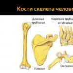
- Long or tubular. Such elements have a middle cylindrical part with a cavity inside and two wide ends, which are covered with a thick layer of cartilage (for example, human leg bones).
- Wide. These are the pectoral and pelvic, as well as the bones of the skull.
- Short. Such elements are characterized by irregular, multifaceted and rounded shapes (for example, wrist bones, vertebrae, etc.).

How are they connected?
The human skeleton (we will see the name of the bones below) is a set of separate bones that are connected to each other. The order of these elements depends on their immediate function. Distinguish between discontinuous and continuous connection of human bones. Let's consider them in more detail.
Continuous connections. These include:
- Fibrous. The bones of the human body are interconnected by a dense connective tissue pad.
- Bone (that is, the bone is completely healed).
- Cartilaginous (intervertebral discs).
Discontinuous connections. These include synovial, that is, between the articulating parts there is an articular cavity. Bones are held in place by a closed capsule and the muscle tissue and ligaments that support it.
Thanks to these features, the arms, bones of the lower extremities and the trunk as a whole are able to set the human body in motion. However, the physical activity of people depends not only on the compounds presented, but also on the nerve endings and bone marrow, which are contained in the cavity of these organs.
Skeleton functions
In addition to the mechanical functions that support the shape of the human body, the skeleton provides the ability to move and protect the internal organs. In addition, it is a place of hematopoiesis. Thus, new blood cells are formed in the bone marrow.
Among other things, the skeleton is a kind of storage for most of the body's phosphorus and calcium. That is why it plays an essential role in the metabolism of minerals.
Human Skeleton with Bones Name
The adult skeleton is made up of about 200+ elements. Moreover, each part of it (head, arms, legs, etc.) includes several types of bones. It should be noted that their name and physical characteristics vary considerably.
Head bones
The human skull has 29 parts. Moreover, each section of the head includes only certain bones:
1. The brain department, consisting of eight elements:

2. The facial section consists of fifteen bones:
- palatine bone (2 pcs.);
- opener;
- (2 pcs.);
- upper jaw (2 pcs.);
- nasal bone (2 pcs.);
- lower jaw;
- lacrimal bone (2 pcs.);
- lower nasal concha (2 pcs.);
- hyoid bone.
3. Bones of the middle ear:
- hammer (2 pcs.);
- anvil (2 pcs.);
- stirrup (2 pcs.).
Torso
Human bones, whose names almost always correspond to their location or appearance, are the most easily examined organs. So, various fractures or other pathologies are quickly detected using a diagnostic method such as radiography. It should be especially noted that some of the largest human bones are the bones of the body. This includes the entire vertebral column, which consists of 32-34 individual vertebrae. Depending on the functions and location, they are divided:
- thoracic vertebrae (12 pcs.);
- cervical (7 pcs.), including epistrophy and atlas;
- lumbar (5 pcs.).
In addition, the bones of the trunk include the sacrum, coccyx, rib cage, ribs (12 × 2) and sternum.
All these elements of the skeleton are designed to protect the internal organs from possible external influences (bruises, blows, punctures, etc.). It should also be noted that in the case of fractures, the sharp ends of the bones can easily damage the soft tissues of the body, which will lead to severe internal hemorrhage, which is most often fatal. In addition, it takes much longer for such organs to grow together than for those located in the lower or upper extremities.
Upper limbs
The bones of the human hand include the largest number of small elements. Thanks to such a skeleton of the upper limbs, people are able to create household items, use them, and so on. Like the spinal column, a person's hands are also subdivided into several sections:

- Shoulder - humerus (2 pieces).
- Forearm - ulna (2 pieces) and radius (2 pieces).
- A brush that includes:
- the wrist (8 × 2), consisting of the scaphoid, lunate, triangular and pisiform bones, as well as the trapezoid, trapezius, capitate and hook-shaped bones;
- the metacarpus, consisting of the metacarpal bone (5 × 2);
- the bones of the fingers (14 × 2), consisting of three phalanges (proximal, middle and distal) in each finger (except for the thumb, which has 2 phalanges).
All the presented human bones, the names of which are quite difficult to remember, allow you to develop hand motor skills and perform the simplest movements that are extremely necessary in everyday life.
It should be especially noted that the constituent elements of the upper limbs are subject to fractures and other injuries most often. However, such bones grow together faster than others.
Lower limbs

Human leg bones also contain a large number of small elements. Depending on their location and functions, they are divided into the following departments:
- Lower limb belt. This includes the pelvic bone, which consists of the ischium and pubis.
- The free part of the lower limb, consisting of the thighs (femur - 2 pieces; patella - 2 pieces).
- Shin. Consists of the tibia (2 pieces) and the fibula (2 pieces).
- Foot.
- Tarsus (7 × 2). It consists of two bones each: calcaneal, ram, scaphoid, medial wedge-shaped, intermediate wedge-shaped, lateral wedge-shaped, cuboid.
- Metatarsus, consisting of the metatarsal bones (5 × 2).
- Finger bones (14 × 2). Let's list them: middle phalanx (4 × 2), proximal phalanx (5 × 2) and distal phalanx (5 × 2).
Most common bone disease
Experts have long established that it is osteoporosis. It is this deviation that most often causes sudden fractures, as well as pain. The unofficial name of the presented disease sounds like "quiet thief". This is due to the fact that the disease proceeds imperceptibly and extremely slowly. Calcium is gradually washed out of the bones, which entails a decrease in their density. By the way, osteoporosis often occurs in old or mature age.
Aging bones
As mentioned above, in old age, the human skeletal system undergoes significant changes. On the one hand, bone loss begins and the number of bone plates decreases (which leads to the development of osteoporosis), and on the other hand, excessive formations appear in the form of bone growths (or so-called osteophytes). Calcification of the articular ligaments, tendons and cartilage also occurs at the site of their attachment to these organs.
Aging of the osteoarticular apparatus can be determined not only by the symptoms of pathology, but thanks to such a diagnostic method as radiography.
What changes occur as a result of bone atrophy? Such pathological conditions include:
- Deformation of the articular heads (or the so-called disappearance of their rounded shape, grinding of the edges and the appearance of corresponding angles).
- Osteoporosis. When examined on an X-ray, the bone of a sick person looks more transparent than that of a healthy one.
It should also be noted that patients often show changes in bone joints due to excessive lime deposition in the adjacent cartilaginous and connective tissue tissues. As a rule, such deviations are accompanied by:
- Narrowing of the articular x-ray gap. This occurs due to calcification of the articular cartilage.
- Strengthening the relief of the diaphysis. This pathological condition is accompanied by calcification of the tendons at the site of bone attachment.
- Bone growths, or osteophytes. This disease is formed due to calcification of the ligaments at the site of their attachment to the bone. It should be especially noted that such changes are especially well detected in the hand and spine. In the rest of the skeleton, there are 3 main X-ray signs of aging. These include osteoporosis, narrowing of joint spaces and increased bone relief.
In some people, such symptoms of aging may appear early (at about 30-45 years old), while in others - late (at 65-70 years old) or not at all. All the described changes are quite logical normal manifestations of the activity of the skeletal system at an older age.

- Few people know, but the hyoid bone is the only bone in the human body that has nothing to do with others. Topographically, it is located on the neck. However, it is traditionally referred to as the facial region of the skull. Thus, the sublingual element of the skeleton with the help of muscle tissue is suspended from its bones and connected to the larynx.
- The longest and strongest bone in the skeleton is the femur.
- The smallest bone in the human skeleton is found in the middle ear.
We continue to delve into the anatomy, this time we will tell the children about the human skeleton. Difficult topics need to be presented to the child in interesting activities. Initially, we will pay attention if interest in our own body is already present, then we will analyze what exactly your little student likes: experiments, modeling from plasticine, application - everything can be used. In the article, I share the complete information on lessons on this topic with my son.
- Human skeleton for younger preschoolers
- Human Skeleton With Bones Name - Cards
- The structure of the human skeleton: head, torso, limbs
Hello dear readers, I welcome you to the blog. Today we will have a fascinating journey into the world of human bones. That's right, we will try, like cartoon characters, to delve into the bowels of the body. It is up to you to decide whether we will travel on a magic bus or a flying ship. The main thing is that our little passengers are interested. Go!
This is the first crossword puzzle in the son's life in his 5 years 6 months. For my child's knowledge, it turned out to be easy enough, which testifies to the full assimilation of information from children's encyclopedias. I will mention the literature of our children's library in the course of the story.
I wrote questions by hand on 6 cards, and on a separate sheet I drew a grid to fill out. You can do the same if you want, but first assess your child's knowledge. If the answers to the questions are not yet familiar to him, postpone this crossword puzzle until the end of the necessary topics.


Questions:
- Not a clock, but ticking.
- The train is endlessly delivering nutrients to the body.
- When full, he is silent. When hungry - hums.
- The organ of vision.
- Respiratory organ of a person.
- He speaks and eats.
Alexander gladly got down to business, he was really interested in solving the crossword puzzle. After graduation, I was ordered a new one about plants and their cultivation.


Most likely your child became interested in his own body during his early preschool years. After all, kids are so curious and start asking a lot of questions. But do not rush and take the child on an excursion to the medical institute, limit yourself to examining such a skeleton of a person from a book My body from head to toe... Where the girl Anya talks about human bones, our muscles and how it grows.

If the child's things, from which he grew up, have survived, then take them out and talk about how his body is changing. Will the baby guess that the size of shoes and clothes changes due to the fact that his bones grow? After reading this book, you will definitely guess! At this stage, it will be a great addition to assemble your skeleton, even a 5-year-old child will cope with it.
Many have preserved X-ray pictures at home, show them to your little student. Consider together and let it be possible to guess which part of the skeleton is in the picture. If they are of good quality, you can even see the texture of the bones. We had a snapshot of Alexander's ribs at the age of three and his mother's foot.

For children from the age of four, the book "Secrets of a Man" from the Magic Doors series will be interesting and understandable. It already provides information on anatomy, but still in a form that is easy for children to understand.
 Increase
 It was thanks to this book that we decided to fool around and paint our skeleton. The advantages of such games are that the child feels each of his bones while drawing, and then can see himself in the mirror. My skeleton then demanded to draw the pelvic bone, but we will not show you that.

I cannot but mention the book of the publishing house MYTH “Bones and Skeletons”, where the kid can see the human skeleton at his own height, as well as examine the skeletons of different animals.
Show the children the human skeleton in a video that is not very animated, but still better perceived than a slide presentation.
Skeleton. Body structure for children - educational cartoon
You can also watch cartoons about Adiba, who we already know from. Adibu travels on the skeleton "Why I stand up straight":
And the explanation about human muscles “Why I move”:
For little fans of educational cards, there are wonderful manuals that include a human skeleton with the name of bones. They appeared with us a long time ago in Russian, English, French and Spanish. Two lovely mothers Katrin and Olga shared them with everyone, here you can download the cards. As you can see in the photo, we are talking not only about the human skeleton with the name of bones, but also the name of all muscles and organs.


I strongly advise you to immediately laminate the cards, as they will be useful to you not only in introductory classes in anatomy, but also in the study of foreign languages. We do not live in Russia, so this is very important in our case. After all, there is nothing worse when you want to tell what you know and cannot because of ignorance of the terms in the interlocutor's language.
The structure of the human skeleton
So let's move on to more serious knowledge. The first thing we will explain to the child is that the human skeleton is divided into the following parts:
- Head skeleton;
- torso;
- upper limbs (shoulder girdle, limbs);
- lower limbs (pelvic girdle, limbs).
If you show this in a picture or on a skeleton model, then the preschooler will definitely understand.


Human head skeleton
The skeleton of a human head is a skull, our children will learn about this from cartoons long before we decide to tell them about their own body. It will be enough for a preschool child to know that the skull reliably protects his brain, which in turn is very soft and vulnerable.
Also, many children may be interested in why there is no nose on the skull? We explain that in fact the nose consists of soft cartilage adhered to the bone. And after death, the cartilage decomposes.
Let's take a look at the skeleton diagram in the book The human body... What will the child immediately notice in the skull?
 Photo enlarges when clicked
 - Eye sockets that protect our eyes;
- teeth fixed by roots in the upper and lower jaws;
- the back of the skull is shorter than the front.
Explain that our brain is located in the back. The only movable part of the skull is the lower jaw. Let the child open and close his mouth, he himself will feel it.
If you want to go deeper, then take apart some of the bones of the skull, which are not very different from the words familiar to the child. Show on your head, and let him repeat after you, show on his.
- The forehead is the frontal bone.
- The temples are the temporal bone.
- The nose is the nasal bone.
- The occiput is the occipital bone.
- The crown is the parietal bone.
- Cheekbones - Zygomatic bone.
- The lower jaw is the mandibular bone.
- The upper jaw is the maxillary bone.
Since the lesson is designed for preschoolers, it is enough for them to explain that the skeleton of the torso consists of the spine and chest... Ribs protect the heart and lungs, and a person has 12 pairs of ribs in total. If the child already knows how to count, then it will not be difficult for him to add 12 + 12 and find out the total number.


The spine is our main support that supports our head and torso. In addition, it protects the spinal cord located inside. In the spine, between the small bones, there are intervertebral discs, they are hard but mobile. It is they who allow us to bend.
Let's do an experiment! What gives us the ability to be flexible?
As we learned, the spine is made up of many small bones. Between them there are gaps of solid, but moving areas. Let's take a look at how this happens.
We need:
- Chenille wire;
- 2 ballpoint pens;
- hacksaw.
We take out all the details of ballpoint pens, we only need a frame (plastic tube). We leave one tube as it is, it should have open holes on both sides. Saw the other into pieces.
First, we ask the child to put the whole tube on the chenille wire and bend it slightly. Does not work? This is how our spine, if it consisted of a solid bone, we would not be able to bend, bend to the sides, many games and movements would be inaccessible to us.
Now we ask the child to put on pieces of plastic tubing, leaving gaps like intervertebral discs. Well, how now, our "spine" has become more flexible?

After this experiment, ask your child to make different body movements. Let him concentrate on the spine, feel its flexibility.

The functions of a person's limbs - arms and legs - are completely different. The legs are responsible for support and movement. And the hands provide a variety of complex movements. We ask the child to take objects with their feet and be like their hands, it is fun and he will immediately understand the difference in functions. The skeleton of the hand consists of 27 bones, and the skeleton of the foot consists of 26 bones.


Alexander and I disassembled only one limb in detail, my son made it out of plasticine.

Observing the child's work, I realized that any knowledge of the human skeleton can be well understood and learned by doing such plasticine X-rays. Indeed, during the creation of such a layout, you have to analyze, count the number of parts, pay attention to their shape.
So how many bones are in a human skeleton?
The adult skeleton consists of 200-218 bones. And the skeleton of a newborn is about 300. What happens then? The baby develops and some bones grow together, larger bones are formed from them. Men and women do not differ in the number of bones - dad and mom can have the same number of bones.
Dear parents, different sources provide information about the skeleton of an adult with 206 bones, 210, a little more than 200. And all these data are correct. Just explain to the child that each organism is individual, the fusion of children's bones is different for everyone. So the data 200-218 is optimal.
- Our skull consists of 29 bones.
- Torso skeleton:
The vertebral column consists of 32-34 vertebrae;
The rib cage consists of 37 bones, which includes 12 pairs of ribs. - Upper limb bones 80.
- Lower limb bones 60.
The total count is as follows: 29 + 37 + 80 + 60 = 206. That is why many sources give this figure, but do not forget about individuality.
How much does a human skeleton weigh?
We all know the expression “light bones and heavy bones”. Sometimes you take a child in your arms and wonder how light or heavy it is - the appearance is sometimes deceiving. Despite this, there is a table according to which it is customary to calculate the weight of a human skeleton:
The bones of a man make up 17-18% of the body weight.
Women - 16% of the total weight.
The weight of a child's skeleton is 14% of the weight of a child.
If there is a scale at home, then weigh yourself with the whole family and calculate the weight of the bones of mom, dad, child. This presentation of information will surely be remembered by the child.
Now, after all that has been passed, you can watch the video of the Human Skeleton, to consolidate knowledge.
Even though bones are very light, they are also very strong. But how strong they are depends on how much calcium carbonate they contain. Let's do an experiment!
What we need:
- Dried, clean chicken bone (leg or wing bone, we have both);
- cones for the experiment (glass);
- white vinegar (we have 5%).


We give the child a bone and ask him to try to break it. We note how tough it is and does not lend itself to children's hands. We examine the bone under a magnifying glass and from the sides we can clearly see the spongy bone tissue.


Now we place the chicken bones in flasks, we have three of them, and cover with vinegar.


Let the bones soak in the vinegar for 1-3 days, then pour out the vinegar. We got the first bone from the winglet, the thinnest one, in a day. Now let your child touch the bone and see the difference. It can be seen how the edges of the bone bend. The child is impressed!


We took out the second and third bones in three days. If you want more effect, you can drain and renew the vinegar once a day. And you can take vinegar essence, but we don't sell such miracles. The bone from the winglet, after 3 days, really bent perfectly along its entire length. But the thick bone from the leg softened only at the edges. Now you can easily break open and see the inside of the medullary canal.


Conclusions of the experiment
Bones are made from calcium carbonate and a soft collagen material. When the chicken bone was placed in a glass of vinegar, the acetic acid dissolved the calcium carbonate and almost only collagen remained. Calcium is needed to make our bones strong. The composition of our bones changes depending on what we eat (the composition of the food). Several foods that are high in calcium include milk, cheese, soy products, beans, almonds, fish (in canned food), and cabbage. After such a lesson, the child understands how important their use is.
On the topic of what human bones are made of, Alexander watched a cartoon that sunk into his soul. I asked for a review for three days. In my opinion, for preschoolers, the topic is well disclosed, but difficult. The child's opinion suggests otherwise. After the screenings, the son can take an anatomy exam on leukocytes and blood cells.
- And what would a person be without bones?
I asked Alexander such a provocative question. My baby lay down on the floor and began to move like a slug.
- Like a puddle of leather!
Yes, this is the comparison made by my boy. And I invited him to see it clearly. Since a puddle, then water. I took a rubber glove, poured water from the tap into it - and so we got a brush without bones!


Dear friends, our journey through the human skeleton is over. Finally, I'll show you what gift my son decided to give me for my birthday, which coincided with our lessons. He asked me not to spy on, so that I would get a real surprise. And here he is!


- Look mom, the skull is smiling at you! - with these words I was presented with a gift.
And I'm sure that no mother has received such a wonderful human skeleton on her birthday.
